 뒤로
Axial Skeleton: Structure, Classification, and Bone Markings
스터디 가이드 - 스마트 노트
 자료에 맞춘 맞춤형 노트, 핵심 정의, 예시, 맥락을 확장해 제공합니다.
Axial Skeleton
Overview and Components
The axial skeleton forms the central axis of the human body and is essential for protection and support of vital organs. It consists of 80 bones, including the skull, vertebral column, thorax, hyoid bone, and auditory ossicles.
Skull: Composed of cranial and facial bones.
Hyoid Bone: Located in the neck, not articulating with other bones.
Auditory Ossicles: Three small bones in each ear.
Vertebral Column: Supports the trunk and protects the spinal cord.
Thorax: Includes the sternum and ribs, protecting thoracic organs.
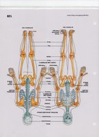
Classification of Bone
Bone Types and Examples
Bones are classified by their shapes, which relate to their functions and structural properties.
Long Bones: Longer than they are wide; found in arms, legs, and phalanges.
Short Bones: Nearly equal in length and width; found in carpals (wrist) and tarsals (ankle).
Flat Bones: Thin and broad; found in the sternum, ribs, and skull.
Irregular Bones: Complex shapes; found in vertebrae, some skull bones, and hipbones.
Sesamoid Bones: Embedded in tendons; example: kneecap (patella).
Sutural Bones: Small bones between cranial bones (not always present).
Bone Surface Markings
Projections, Depressions, and Openings
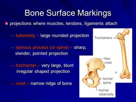
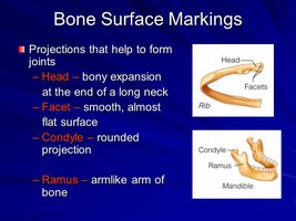
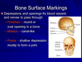
Bone surfaces are rarely smooth; they feature various markings that serve as attachment points for muscles, tendons, and ligaments, or as passageways for nerves and blood vessels.
Projections: Tuberosity, spinous process, trochanter, crest.
Joint-forming projections: Head, facet, condyle, ramus.
Depressions and Openings: Foramen, meatus, fossa.
The Skull
Cranial and Facial Bones
The skull is the most complex structure in the axial skeleton, formed from two sets of bones:
Cranial Bones (8): Protect the brain and provide attachment sites for head muscles.
Facial Bones (14): Form the face, hold the eyes, provide cavities for taste and smell, secure teeth, and anchor facial muscles.
All skull bones are united by immobile interlocking joints called sutures.
Coronal suture: Between parietal and frontal bones.
Sagittal suture: Between two parietal bones.
Lambdoid suture: Between parietal and occipital bones.
Squamous suture: Between parietal and temporal bones.
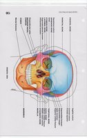
Skull Views
Different views of the skull are necessary for studying its structure and bone markings. 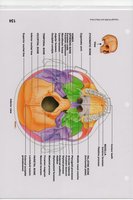
Fontanelles
Newborn Skull Features
At birth, the skull contains soft spots called fontanelles, which are dense connective tissue regions that allow for growth and flexibility during birth.
Anterior Fontanelle: Largest, ossifies at 18-24 months.
Posterior Fontanelle: Ossifies at 2 months.
Sphenoidal Fontanelle: Ossifies at 3 months.
Mastoid Fontanelle: Ossifies at 1-2 months.
Paranasal Sinuses
Structure and Function
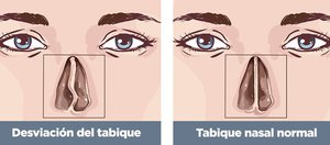
Paranasal sinuses are cavities within facial and cranial bones, lined with mucous membranes and continuous with the nasal cavity. They act as resonating chambers to enhance voice quality.
Frontal sinus
Ethmoidal sinus
Sphenoidal sinus
Maxillary sinus
Hyoid Bone
Location and Function
The hyoid bone is unique as it does not articulate with any other bone. It is suspended by ligaments and muscles from the styloid processes of the temporal bone.
Supports the tongue.
Serves as an attachment for vocal cords and laryngeal cartilages.
Fracture of the hyoid bone is associated with strangulation.
Eye Orbit
Structure
The eye orbit is formed by several bones, moving inward from the nasal bone. 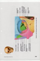
Sphenoid Bone
Location and Significance
The sphenoid bone is a central bone of the skull, housing the sella turcica, which holds the pituitary gland. 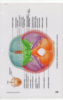
The Mandible
Structure and Bone Markings
The mandible is the lower jawbone and features several important bone markings, including the condylar process, mandibular foramen, and mental foramen. 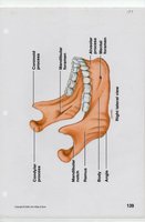
Middle Ear Ossicles
Function
The middle ear contains three small bones:
Malleus
Incus
Stapes
These ossicles transmit sound vibrations from the eardrum to the inner ear, where they are converted into nerve impulses.
Summary Table: Cranial Bones and Markings
Comparison of Cranial Bones and Their Notable Structures
Bone | Markings/Structures |
|---|---|
Frontal | Supraorbital foramen, coronal suture |
Parietal | Sagittal suture |
Occipital | Foramen magnum, occipital condyles, lambdoid suture |
Temporal | Zygomatic process, mandibular fossa, external auditory meatus, styloid process, mastoid process, squamous suture |
Sphenoidal | Sella turcica |
Ethmoid | Crista galli, cribriform plate |
Additional info:
Bone markings are critical for understanding muscle attachment, joint formation, and passage of neurovascular structures.
Fontanelles are clinically important for assessing infant development and hydration status.
Paranasal sinus inflammation (sinusitis) can result from infection, allergies, or anatomical deviations such as a deviated nasal septum.
