 뒤로
Axial Skeleton: Structure, Function, and Key Components
스터디 가이드 - 스마트 노트
 자료에 맞춘 맞춤형 노트, 핵심 정의, 예시, 맥락을 확장해 제공합니다.
Axial Skeleton and Skeletal System Overview
Divisions of the Skeletal System
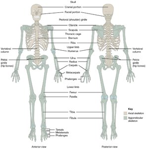
The skeletal system is the rigid internal framework of the body, consisting of bones, cartilages, and ligaments. It is divided into two main parts: the axial skeleton and the appendicular skeleton.
Axial Skeleton: Composed of 80 bones, it protects the brain, spinal cord, heart, and lungs. It includes the skull, vertebral column, and thoracic cage.
Appendicular Skeleton: Composed of 126 bones, it facilitates movement of the limbs and includes the bones of the upper and lower limbs, pectoral girdle, and pelvic girdle.
Key Functions:
Supports body weight and allows for movement
Protects internal organs
Stores calcium, phosphate, and fat
Red bone marrow produces blood cells
Skull: Cranium and Facial Bones
Cranial Bones
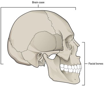
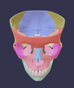
The cranium consists of several bones fused together to protect the brain and house ear structures. The main cranial bones are:
Frontal bone: Forehead and upper part of orbits
Parietal bones: Top of the cranium
Temporal bones: Sides and floor of the cranium
Ethmoid bone: Forms part of the cranial floor and nasal cavity
Sphenoid bone: Central cranial floor, anchors other cranial bones
Occipital bone: Posterior skull, forms joint with the atlas (C1)
Facial Bones
Facial bones support the face, form the nasal cavity, enclose the eyeballs, and support the teeth. Key facial bones include:
Lacrimal bones: Medial orbit wall, contains tear duct groove
Nasal bones: Bridge of the nose
Maxilla: Upper jaw, orbital floors, roof of mouth
Mandible: Lower jaw, only movable skull bone
Zygomatic bones: Cheekbones
Palatine bones: Hard palate and lateral nasal cavity
Vomer: Nasal septum
Inferior nasal conchae: Lateral nasal cavity wall

Paranasal Sinuses
Paranasal sinuses are air-filled spaces within certain skull bones, connected to the nasal cavity and lined with mucosa. They lighten the skull and add resonance to the voice.
Frontal sinus: Above the eyebrows
Sphenoid sinus: Within sphenoid bone
Ethmoid air cells: Lateral sides of ethmoid bone
Maxillary sinus: Largest, within maxillary bones
Muscles of Facial Expression and Mastication
Facial Expression Muscles
Muscles that create facial expressions originate on the skull and insert into connective tissue and dermis. Major muscles include:
Occipitofrontalis: Moves scalp and eyebrows
Orbicularis oculi: Closes the eye
Orbicularis oris: Moves the lips
Buccinator: Compresses the cheek
Corrugator supercilii: Moves the eyebrows
Muscles of Mastication (Chewing)
These muscles move the mandible for biting and chewing:
Masseter: Elevates mandible
Temporalis: Elevates and retracts mandible
Buccinator: Keeps food in place
Lateral pterygoid: Depresses and protracts mandible
Medial pterygoid: Elevates and protracts mandible

Hyoid Bone and Associated Muscles
Hyoid Bone
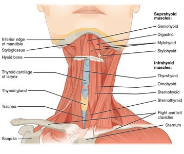
The hyoid bone is a small, U-shaped bone in the upper neck, not directly attached to other bones. It supports muscles involved in swallowing and speech.
Suprahyoid muscles: Raise hyoid, floor of mouth, and larynx
Infrahyoid muscles: Depress hyoid and control larynx position

Vertebral Column
Structure and Regions
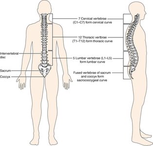
The vertebral column is a flexible structure composed of vertebrae separated by intervertebral discs. It supports the head, neck, and body, and protects the spinal cord.
Cervical vertebrae (C1–C7): Neck
Thoracic vertebrae (T1–T12): Chest, attach to ribs
Lumbar vertebrae (L1–L5): Lower back
Sacral vertebrae: Fused to form pelvis
Coccygeal vertebrae: Fused to form tailbone

Curvatures of the Vertebral Column
The vertebral column has four curvatures that increase strength and flexibility:
Primary curves: Thoracic and sacrococcygeal (present at birth)
Secondary curves: Cervical (develops when infant holds head up) and lumbar (develops when child stands/walks)
Structure of a Typical Vertebra
Each vertebra has a body, vertebral arch, vertebral foramen, and processes for muscle and ligament attachment.
Body: Supports weight
Vertebral arch: Protects spinal cord
Vertebral foramen: Passage for spinal cord
Intervertebral foramen: Passage for spinal nerves
Processes: Transverse, spinous, superior/inferior articular
Intervertebral Discs and Ligaments
Intervertebral discs are fibrocartilaginous pads between vertebrae, consisting of anulus fibrosus (outer layer) and nucleus pulposus (inner gel-like core). Ligaments stabilize the vertebral column.
Anterior longitudinal ligament: Prevents excessive backward bending
Supraspinous ligament: Supports column during forward bending
Nuchal ligament: Supports skull
Posterior longitudinal ligament and ligamentum flavum: Support column during forward bending

Cervical Vertebrae: Atlas and Axis
The first two cervical vertebrae (C1 and C2) are specialized for head movement:
Atlas (C1): Supports the skull, allows "yes" motion
Axis (C2): Has the dens (odontoid process), allows "no" motion


Thoracic Vertebrae
Thoracic vertebrae have larger bodies, long downward spinous processes, and facets for rib articulation.
Costal facets: Articulate with rib heads
Articular facet on transverse process: Articulates with rib tubercle

Thoracic Cage (Rib Cage)
Structure and Function
The thoracic cage consists of 12 pairs of ribs, costal cartilages, and the sternum. It protects the heart and lungs.
Sternum: Manubrium (superior), body (central), xiphoid process (inferior)
Ribs: True ribs (1-7, direct attachment), false ribs (8-12, indirect or no attachment), floating ribs (11-12)
Parts of a Typical Rib
Head: Articulates with vertebral body
Neck: Narrowed portion
Tubercle: Articulates with transverse process
Body (shaft): Main part of rib
Embryonic Development and Fontanels
Development of the Axial Skeleton
The axial skeleton forms during embryonic development via intramembranous and endochondral ossification. Fontanels (soft spots) allow for skull molding during birth and rapid brain growth.
Fontanels: Anterior, posterior, sphenoidal, mastoid
Sutures: Interlocking projections between skull bones
Summary Table: Axial Skeleton Components
Component | Main Function | Key Features |
|---|---|---|
Skull | Protects brain, supports face | Cranial and facial bones, paranasal sinuses |
Hyoid bone | Supports tongue, aids swallowing | U-shaped, not attached to other bones |
Vertebral column | Supports body, protects spinal cord | Cervical, thoracic, lumbar, sacral, coccygeal regions |
Thoracic cage | Protects heart and lungs | Ribs, sternum, costal cartilages |
