 뒤로
Bacterial Structure, Growth, and Pathogenesis: Study Notes for Anatomy & Physiology
스터디 가이드 - 스마트 노트
 자료에 맞춘 맞춤형 노트, 핵심 정의, 예시, 맥락을 확장해 제공합니다.
Bacterial Growth and Reproduction
Binary Fission
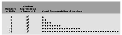
Binary fission is the primary method of reproduction in bacteria, resulting in exponential population growth. This process is asexual and involves the division of a single cell into two genetically identical daughter cells. - Generation time: The time required for a bacterial cell to divide, which can range from 20 minutes to 20 hours depending on the species and environmental conditions. - Exponential growth: Bacterial populations double with each generation, following the formula , where is the final number of cells, is the initial number, and is the number of generations. 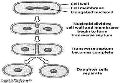
Bacterial Morphology
Bacterial Shapes
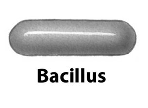
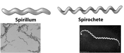
Bacteria exhibit a variety of shapes, which are important for identification and classification. - Coccus (pl. cocci): Spherical shape. 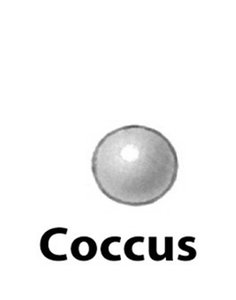 - Bacillus (pl. bacilli): Rod-shaped.
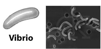
 - Spiral forms: Includes vibrio (curved rod), spirillum (rigid spiral), and spirochete (flexible spiral).
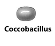
 - Coccobacillus: Short, oval rod.
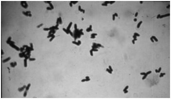
 - Pleomorphic: Variable shapes, often seen in certain bacterial species.
Bacterial Cell Arrangements
Bacterial cells can be arranged in characteristic patterns based on their division. - Diplo-: Pairs (e.g., diplococcus). - Strepto-: Chains (e.g., streptococcus, streptobacillus). 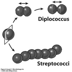 - Staphylo-: Clusters (e.g., staphylococcus).
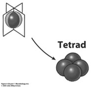
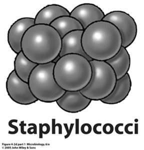 - Tetrad: Groups of four cells.
Prokaryotic Cell Structure
General Structure
Prokaryotic cells (bacteria) have a simple structure compared to eukaryotes, lacking membrane-bound organelles. - Cell wall: Provides structural support and protection. - Cell membrane: Regulates transport of substances in and out of the cell. - Cytoplasm: Contains water, enzymes, and other molecules. - Ribosomes: Sites of protein synthesis. - Nucleoid: Region containing the bacterial chromosome (DNA). - Plasmids: Small, circular DNA molecules carrying nonessential genes. - Flagella: Used for motility. - Pili (fimbriae): Used for attachment and conjugation. - Capsule/slime layer (glycocalyx): External polysaccharide layer for protection and attachment. 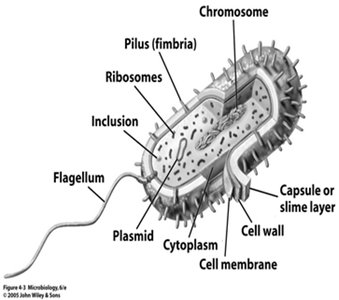
Bacterial Cell Wall Types
Peptidoglycan Structure
Peptidoglycan is a complex polymer forming the bacterial cell wall, providing rigidity and shape. - Structure: Composed of sugar chains (N-acetylglucosamine and N-acetylmuramic acid) cross-linked by peptides. 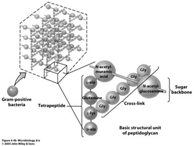
Gram-Positive Bacteria
- Thick peptidoglycan layer: Up to 40 layers, retains stains in Gram staining. - Teichoic acids: Present in cell wall, contribute to cell wall stability. 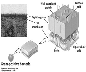
Gram-Negative Bacteria
- Thin peptidoglycan layer: Located between inner cell membrane and outer membrane. - Outer membrane: Contains lipopolysaccharides, provides additional protection. - Does not retain Gram stain: Appears red or pink after staining. 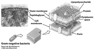
Acid-Fast Bacteria
- Less peptidoglycan: Surrounded by a thick, waxy lipid layer. - Slow growth: Due to reduced permeability. - Examples: Mycobacterium species (tuberculosis, leprosy). 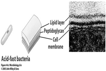
Bacteria Without Cell Walls
- Example: Mycoplasma species, cause "walking pneumonia." 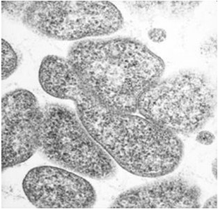
Gram Stain Technique
The Gram stain is a differential staining method used to distinguish between Gram-positive and Gram-negative bacteria. - Gram-positive: Stain purple or blue. - Gram-negative: Stain red or pink. 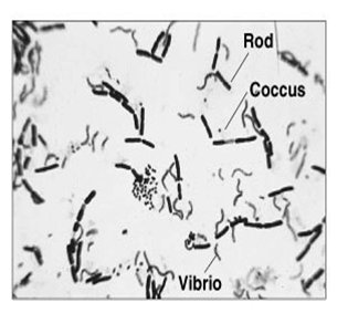
Control of Bacteria by Cell Wall Damage
- Penicillin: Inhibits peptidoglycan synthesis, effective against Gram-positive bacteria. - Lysozyme: Enzyme found in tears, sweat, and saliva; digests peptidoglycan.
Internal Cell Structures
Cytoplasm
The cytoplasm is the internal fluid of the cell, mainly composed of water, enzymes, and nutrients. 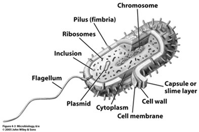
Ribosomes
Ribosomes are the sites of protein synthesis in bacteria. 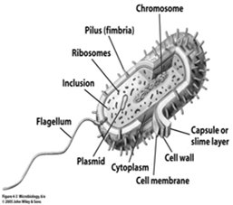
Nucleoid
The nucleoid is the region where the bacterial chromosome (DNA) is located. 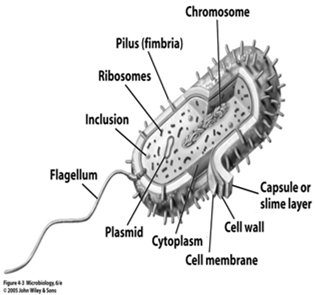
Plasmids
Plasmids are small, circular DNA molecules that carry nonessential genes, often conferring antibiotic resistance. 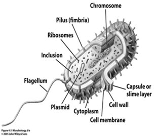
Endospores
Endospores are highly resistant, dormant structures formed by some bacteria under adverse conditions. - Vegetative cells: Metabolically active cells. - Endospores: Dormant stage, survive after vegetative cell dies. - Resistance: Endospores are resistant to drying, heat, radiation, and chemicals. - Examples: Bacillus and Clostridium species (tetanus, botulism, anthrax). 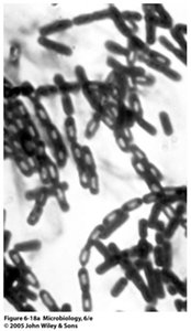
Sporulation and Germination
- Sporulation: Formation of endospore within vegetative cell when conditions are poor. - Germination: Formation of vegetative cell from endospore when conditions improve. 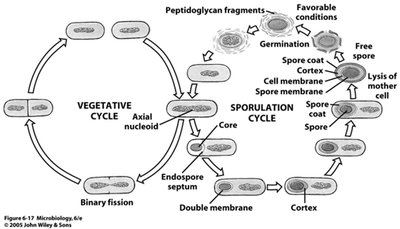
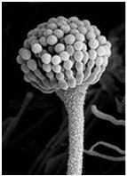
Bacterial Endospores vs. Fungal Spores
Feature | Bacterial Endospores | Fungal Spores |
|---|---|---|
Function | Resistance | Reproduction |
Number Produced | 1 per vegetative cell | Many per mold |
Examples | Bacillus, Clostridium | Aspergillus, Penicillium |
Bacterial Pathogenesis: Anthrax
Etiologic Agent
- Bacillus anthracis: Gram-positive rod, forms endospores.
Reservoir and Transmission
- Reservoir: Soil, disease of cattle. - Transmission: Zoonosis (animal disease spread to humans), occupational exposure (contact with animals, handling wool, hides, meat).
Pathogenesis
- Exotoxins: Cause edema, tissue destruction, bleeding, and kill macrophages.
Forms of Anthrax
- Cutaneous: Spores enter skin wound, 90% of human cases. - Respiratory: Inhalation of spores, bacteria enter bloodstream (septicemia). - Intestinal: Ingestion of spores, damage to intestinal cells.
Symptoms
- Cutaneous: Localized, black scab, mortality rate 10-20% (1% with treatment). - Respiratory: Early fever, cough, rapid progression to septicemia, ~100% mortality rate. - Intestinal: Nausea, bloody diarrhea, ulcerative lesions, 50% mortality rate.
Diagnosis
- Isolation: Organism from clinical specimen, blood tests.
Treatment and Prevention
- Treatment: Antibiotics (ciprofloxacin, doxycycline, amoxicillin), early intervention critical. - Prevention: Vaccine for high-risk groups and livestock, antibiotics if exposure suspected.
External Cell Structures
Flagella
Flagella are whip-like structures used for bacterial motility.
Pili (Fimbriae)
Pili are hair-like appendages used for attachment and DNA transfer. - Attachment pili: Adhere to surfaces (e.g., Neisseria gonorrhoeae to mucous membranes). - Conjugation pili: Join bacteria to transfer DNA (plasmids).
Glycocalyx
The glycocalyx is an external polysaccharide layer, which can be a slime layer (thinner) or capsule (thicker). - Functions: Attachment (e.g., to teeth forming plaque), prevents drying out, protects bacterium from host defenses.
Bacterial Pathogenesis: Pneumococcal Pneumonia
Etiologic Agent
- Streptococcus pneumoniae: Gram-positive diplococcus, has a capsule.
Reservoir and Transmission
- Reservoir: Healthy human carriers. - Transmission: Respiratory droplets, most susceptible are elderly, infants, immunocompromised.
Pathogenesis
- Capsule: Bacteria evade phagocytes due to capsule.
Symptoms
- Early: Mild respiratory symptoms. - Progression: High fever, chills, chest pain, cough, bloody sputum.
Diagnosis
- Sputum sample: Must distinguish from saliva; presence of phagocytes and Gram-positive diplococci.
- Gram stain: Confirms Gram-positive diplococci.
Treatment and Prevention
- Treatment: Antibiotics, but drug-resistant strains are increasing. Untreated cases have a 25% mortality rate. - Prevention: Vaccine (Pneumovax for elderly, Prevnar for children).
Distribution and Other Diseases
- Incidence: 500,000 cases/year in U.S. - Other diseases: S. pneumoniae can cause ear infections, sinusitis, bronchitis, septicemia, and meningitis. Additional info: The notes above expand on the original content with academic context, definitions, and examples to ensure completeness and clarity for Anatomy & Physiology students. Tables and images are included only when directly relevant to the explanation.
