 뒤로
Blood: Structure, Function, and Hemostasis
스터디 가이드 - 스마트 노트
 자료에 맞춘 맞춤형 노트, 핵심 정의, 예시, 맥락을 확장해 제공합니다.
Blood: Structure, Function, and Hemostasis
Functions of Blood
Blood is a vital connective tissue that performs several essential functions in the human body:
Transport: Blood carries oxygen, nutrients, hormones, and waste products throughout the body.
Regulation: It helps maintain fluid and electrolyte balance, acid–base balance, and body temperature.
Protection: Blood defends against infection and contains clotting factors to prevent excessive blood loss.
Composition of Blood
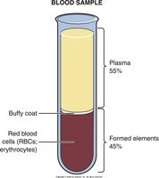
Blood consists of two main components: plasma and formed elements. It is classified as a connective tissue, with cells suspended in a liquid matrix. The color of blood ranges from bright red (oxygenated) to dark blue-red (deoxygenated), depending on oxygen content. The average adult has 4 to 6 liters of blood, with a normal pH of 7.35–7.45 and a viscosity three to five times greater than water.
Plasma: Makes up 55% of blood volume; contains water, proteins, electrolytes, and other solutes.
Formed Elements: Comprise 45% of blood; include erythrocytes (red blood cells), leukocytes (white blood cells), and thrombocytes (platelets).
Origin and Formation of Blood Cells (Hemopoiesis)
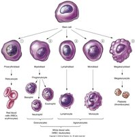
Blood cells are produced through the process of hemopoiesis, which occurs primarily in the red bone marrow. Stem cells differentiate into various blood cell types via myeloid and lymphoid lineages.
Myeloid lineage: Produces erythrocytes, granulocytes, monocytes, and platelets.
Lymphoid lineage: Produces lymphocytes.
Bone Marrow Disorders
Bone marrow can be affected by depression (myelosuppression) or overactivity:
Bone marrow depression: Leads to conditions such as aplastic anemia, leukopenia, and thrombocytopenia.
Bone marrow overactivity: Causes polycythemia vera, resulting in thickened blood, increased cardiac burden, and a ruddy complexion.
Red Blood Cells (Erythrocytes)
Red blood cells are large, round, and doughnut-shaped, losing most organelles as they mature. They produce ATP anaerobically and contain hemoglobin, which consists of globin (protein) and heme (iron-containing substance).
Hemoglobin production: Requires iron, vitamin B12, and folic acid.
Regulation: Erythropoietin, a hormone, regulates RBC production via a negative feedback loop.
Color: Oxygenated blood is bright red; deoxygenated blood is darker.
Removal and Breakdown of Red Blood Cells
Old RBCs are removed by macrophages in the spleen and liver, which phagocytose and recycle their components. Excessive breakdown leads to elevated bilirubin, causing jaundice.
White Blood Cells (Leukocytes)
White blood cells are large, nucleated cells that protect the body by destroying pathogens and removing debris. There are 5,000–10,000 WBCs per microliter of blood, and they migrate to tissues to perform their functions.
Granulocytes: Neutrophils, basophils, eosinophils
Agranulocytes: Lymphocytes, monocytes
Platelets (Thrombocytes)
Platelets are tiny fragments of megakaryocytes, with 150,000–450,000 per microliter of blood. They play a crucial role in preventing blood loss by initiating clot formation.
Blood counts: Complete blood cell count (CBC) and differential WBC count are used to assess blood health.
Hemostasis: Prevention of Blood Loss
Hemostasis is the process that stops bleeding and involves three main steps:
Blood vessel spasm: Constriction of the vessel reduces blood flow.
Formation of a platelet plug: Platelets adhere to the injury site and aggregate.
Blood clotting (coagulation): A cascade of reactions leads to the formation of a fibrin clot.
Clotting Cascade
Stage I: Injury activates clotting factors.
Stage II: In the presence of calcium, platelet chemicals, and PTA, prothrombin is converted to thrombin.
Stage III: Thrombin activates fibrinogen, forming fibrin threads.
Clot retraction and fibrinolysis (clot busting) prevent excessive clot formation. Natural mechanisms and medications help regulate this process.
Blood Types and Compatibility
Blood types are determined by antigens on the RBC membrane and corresponding antibodies in plasma. The ABO system includes types A, B, AB, and O. Antigen–antibody interactions can cause agglutination and hemolysis.
Compatibility: Blood type is determined and cross-matched before transfusion to prevent reactions.
Rh Factor
The Rh classification system is based on the presence or absence of the Rh antigen on RBCs:
Rh-positive (+): RBCs contain the Rh factor.
Rh-negative (−): RBCs lack the Rh factor.
Rh incompatibility can lead to plasma sensitization and conditions such as erythroblastosis fetalis (hemolytic disease of the newborn).
Blood Type | Antigen on RBC | Antibody in Plasma | Can Receive From |
|---|---|---|---|
A | A | Anti-B | A, O |
B | B | Anti-A | B, O |
AB | A and B | None | A, B, AB, O |
O | None | Anti-A, Anti-B | O |
Example: A person with type O blood can only receive type O blood, while a person with type AB can receive any blood type.
Additional info: The notes have been expanded to include definitions, examples, and a summary table for blood types to ensure completeness and academic quality.
