 뒤로
Blood Vessel Structure, Function, and Circulatory Dynamics
스터디 가이드 - 스마트 노트
 자료에 맞춘 맞춤형 노트, 핵심 정의, 예시, 맥락을 확장해 제공합니다.
Blood Vessel Structure and Function
Overview of Blood Vessels
Blood vessels form a closed delivery system that begins and ends at the heart. They are dynamic structures that pulsate, constrict, relax, and multiply, working with the lymphatic system to circulate fluids.
Arteries carry blood away from the heart toward capillaries. Systemic arteries transport oxygenated blood, while pulmonary arteries carry oxygen-poor blood.
Capillaries are exchange vessels, directly serving cellular needs by allowing substances to move across their walls between tissue cells and blood.
Veins carry blood from capillaries toward the heart. Systemic veins carry oxygen-poor blood, while pulmonary veins carry oxygenated blood.
Generalized Structure of Arteries, Veins, and Capillaries
Most blood vessel walls (except capillaries) consist of three layers, or tunics:
Tunica intima: Innermost layer, composed of endothelium and subendothelial layer. Provides a slick surface to reduce friction.
Tunica media: Middle layer, mostly smooth muscle and elastin. Responsible for vasoconstriction and vasodilation, regulating blood flow and pressure.
Tunica externa (adventitia): Outermost layer, mainly collagen fibers, protects and anchors the vessel. Contains vasa vasorum in large vessels.
Capillary wall: Consists only of endothelium with a sparse basal lamina.
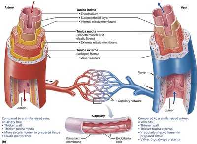
Types of Blood Vessels
Arteries: Pressure Reservoirs, Distributing Vessels, and Resistance Vessels
Arteries are divided into three groups based on size and function:
Elastic arteries: Thick-walled, near the heart (e.g., aorta). Act as pressure reservoirs, expanding and recoiling to maintain continuous blood flow.
Muscular arteries: Distribute blood to specific organs, have the thickest tunica media, and are more vasoactive.
Arterioles: Smallest arteries, control flow into capillary beds via vasodilation and vasoconstriction, and are major determinants of resistance.
Capillaries: Exchange Vessels
Capillaries are the smallest blood vessels, with ultra-thin walls for exchange of gases, nutrients, wastes, and hormones. Most tissues have a rich capillary supply, except poorly vascularized tendons, ligaments, and avascular tissues.
Types of Capillaries
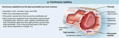
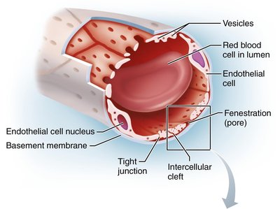
Continuous capillaries: Least permeable, most common, abundant in skin, muscles, lungs, and CNS. Have tight junctions and intercellular clefts for limited passage of fluids and small solutes.
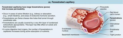
Fenestrated capillaries: Found in areas of active filtration (kidneys), absorption (intestines), and endocrine hormone secretion. Endothelial cells contain pores (fenestrations) for increased permeability.
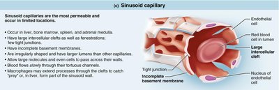
Sinusoidal capillaries: Most permeable, found in liver, bone marrow, spleen, and adrenal medulla. Have large intercellular clefts, fenestrations, and incomplete basement membranes.
Veins: Blood Reservoirs
Veins carry blood toward the heart, gradually thickening from venules to larger veins.
Venules: Formed by uniting capillaries, very porous, allow fluids and WBCs to move into tissues.
Veins: Have all tunics but thinner walls and larger lumens than arteries. Contain up to 65% of blood supply, have adaptations like venous valves to prevent backflow, and venous sinuses with thin walls supported by surrounding tissues.
The Muscular Pump
The muscular pump assists venous return by contracting skeletal muscles around deep veins, "milking" blood toward the heart. 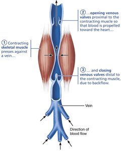
Blood Flow, Pressure, and Resistance
Definitions and Relationships
Blood flow (F): Amount of blood flowing through a vessel, organ, or entire circulation per unit time (ml/min).
Blood pressure (BP): Force per unit area exerted on vessel wall by blood, measured in mm Hg.
Resistance (TPR): Opposition to flow, mainly due to blood viscosity, vessel length, and diameter.
Key Relationships
Blood flow is directly proportional to the pressure gradient () and inversely proportional to total peripheral resistance (TPR):
Resistance varies inversely with the fourth power of vessel radius:
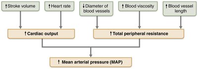
Major Factors That Increase Mean Arterial Pressure (MAP)
Stroke volume, heart rate, diameter of blood vessels, blood viscosity, and vessel length all influence MAP.
Blood Pressure Regulation
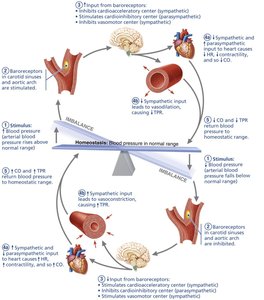
Short-Term Regulation: Neural Controls
Neural controls regulate TPR and CO to maintain MAP.
Baroreceptor reflex: Baroreceptors in carotid sinuses, aortic arch, and large arteries monitor pressure changes and adjust vessel diameter and heart rate.
Chemoreceptor reflexes: Respond to changes in CO2, pH, and O2 levels, influencing cardiovascular center activity.
Higher brain centers: Hypothalamus and cerebral cortex can modify arterial pressure during stress or exercise.
Short-Term Regulation: Hormonal Controls
Hormones regulate MAP via changes in TPR or blood volume.
Epinephrine and norepinephrine: Increase CO and vasoconstriction.
Angiotensin II: Stimulates vasoconstriction and release of aldosterone and ADH.
Atrial natriuretic peptide (ANP): Decreases MAP by promoting vasodilation and salt/water loss.
ADH (vasopressin): Stimulates water conservation and vasoconstriction.
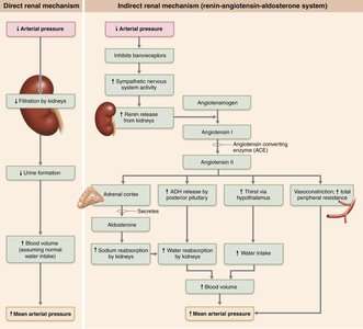
Long-Term Regulation: Renal Mechanisms
Direct renal mechanism: Alters blood volume independently of hormones by adjusting urine output.
Indirect renal mechanism: The renin-angiotensin-aldosterone system increases MAP and ECF volume via hormone release and vasoconstriction.
Summary of Blood Pressure Regulation
The goal is to keep blood pressure high enough for adequate tissue perfusion but not so high as to damage vessels. 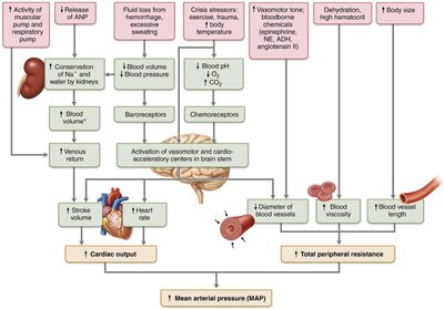
Intrinsic and Extrinsic Controls of Blood Flow
Intrinsic vs. Extrinsic Controls
Intrinsic controls: Local regulation within tissues/organs, using paracrines or muscle properties (autoregulation).
Extrinsic controls: Regulation from outside the tissue/organ, using nerves or hormones to maintain MAP.
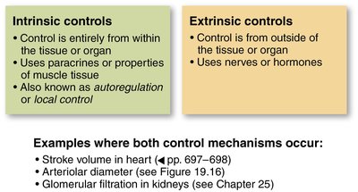
Distribution of Blood Flow During Exercise
Blood flow is redistributed during exercise, with skeletal muscles receiving a greater proportion. 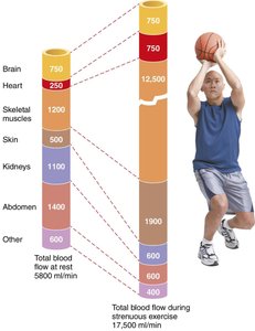
Intrinsic and Extrinsic Control of Arteriolar Smooth Muscle
Metabolic and myogenic mechanisms (intrinsic) regulate local blood flow.
Neural and hormonal mechanisms (extrinsic) maintain MAP and redistribute blood during exercise.
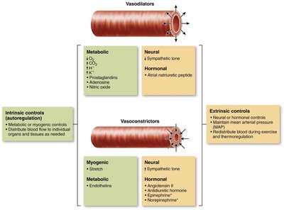
Blood Flow in Special Areas
Skeletal Muscle
Blood flow increases in proportion to metabolic activity during exercise (active hyperemia). 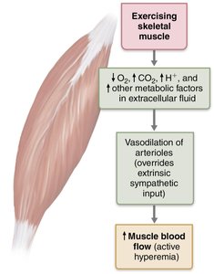
Velocity of Blood Flow
Velocity changes as blood travels through systemic circulation, being fastest in the aorta and slowest in capillaries. 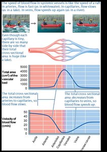
Capillary Exchange and Fluid Movements
Capillary Transport Mechanisms
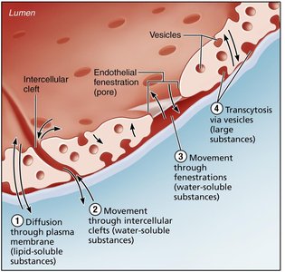
Molecules cross capillaries via four routes:
Diffusion through plasma membrane (lipid-soluble substances)
Movement through intercellular clefts (water-soluble substances)
Movement through fenestrations (water-soluble substances)
Transcytosis via vesicles (large substances)
Bulk Flow Across Capillary Walls
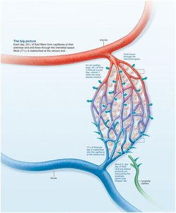
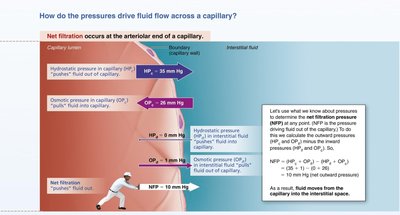
Fluid is forced out of capillaries at the arterial end and returns at the venous end, determined by hydrostatic and osmotic pressures.
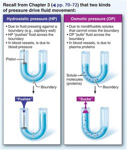
Hydrostatic pressure (HP): Pushes fluid out of capillaries.
Osmotic pressure (OP): Pulls fluid into capillaries.
Net filtration pressure (NFP):
Clinical Homeostatic Imbalances
Varicose Veins
Varicose veins are dilated veins due to incompetent valves, often caused by conditions hindering venous return (e.g., prolonged standing, obesity, pregnancy).
Edema
Edema is an abnormal increase in interstitial fluid volume, resulting from increased capillary hydrostatic pressure, decreased capillary colloid osmotic pressure, or impaired lymphatic drainage.
Circulatory Pathways
Pulmonary and Systemic Circulation
Pulmonary circulation: Short loop from heart to lungs and back.
Systemic circulation: Long loop to all body parts and back.
Major Arteries and Veins
Arteries run deep, veins are both deep and superficial.
Venous pathways are more interconnected.
Brain and digestive systems have unique venous drainage.
Developmental Aspects of Blood Vessels
Endothelium arises from mesodermal cells forming blood islands.
Vascular modifications in fetus include shunts bypassing nonfunctional lungs and liver.
Vessels form throughout life to support tissue growth and healing.
Summary Table: Effects of Selected Hormones on Blood Pressure
Hormone | Effect on BP | Variable Affected | Site of Action |
|---|---|---|---|
Epinephrine/Norepinephrine | Increase | CO (HR, contractility), TPR (vasoconstriction) | Heart (β1), Arterioles (α) |
Angiotensin II | Increase | TPR (vasoconstriction) | Arterioles |
ADH | Increase | TPR (vasoconstriction), Blood volume (water conservation) | Arterioles, Kidney tubules |
Aldosterone | Increase | Blood volume (salt/water conservation) | Kidney tubules |
ANP | Decrease | Blood volume (salt/water loss), TPR (vasodilation) | Kidney tubules, Arterioles |
Key Equations
Additional info:
These notes expand on the original content with academic context, definitions, and examples for clarity and completeness.
