 뒤로
Blood Vessels and the Circulatory System: Structured Study Notes
스터디 가이드 - 스마트 노트
 자료에 맞춘 맞춤형 노트, 핵심 정의, 예시, 맥락을 확장해 제공합니다.
Blood Vessels and the Circulatory System
Overview of Blood Vessels
The circulatory system is composed of a network of blood vessels that transport blood throughout the body. These vessels include arteries, veins, and capillaries, each with distinct structural and functional characteristics.
Arteries: Carry blood away from the heart; typically oxygenated except for the pulmonary arteries.
Veins: Carry blood toward the heart; typically deoxygenated except for the pulmonary veins.
Capillaries: Microscopic vessels where exchange of gases, nutrients, and waste occurs between blood and tissues.
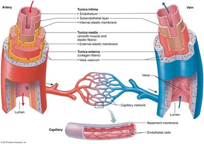
Structural Differences Between Arteries and Veins
Arteries and veins have distinct structural layers that reflect their functions in the circulatory system.
Tunica intima: Innermost layer, composed of endothelium.
Tunica media: Middle layer, contains smooth muscle and elastic fibers; thicker in arteries.
Tunica externa: Outermost layer, composed of connective tissue; provides structural support.
Valves: Present in veins to prevent backflow of blood.
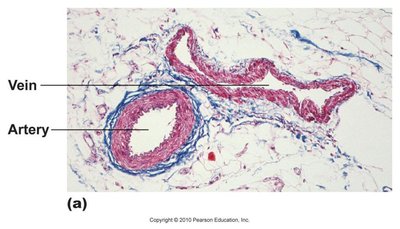
Major Blood Vessel Pathways
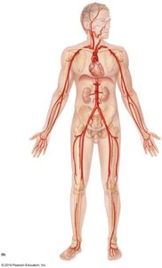
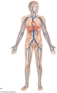
The body contains major arteries and veins that supply and drain blood from various regions. Understanding their pathways is essential for anatomical identification.
Trunks: Short, singular arteries that quickly branch into other major arteries (e.g., pulmonary trunk, brachiocephalic trunk, celiac trunk).
Regional Terms: Axillary, brachial, radial, and ulnar arteries are named based on their anatomical regions.
Cephalic Vessels and the Circle of Willis
The cephalic vessels supply the head and brain. The Circle of Willis is a critical arterial structure providing collateral circulation to the brain.
Circle of Willis: Ensures redundancy in cerebral blood supply; allows compensation for occlusion and equalizes pressure between hemispheres.
Blood Input: Comes from the basilar artery and both internal carotid arteries.
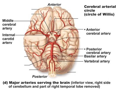
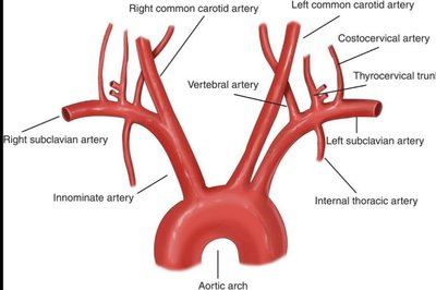
Branches of the Aortic Arch
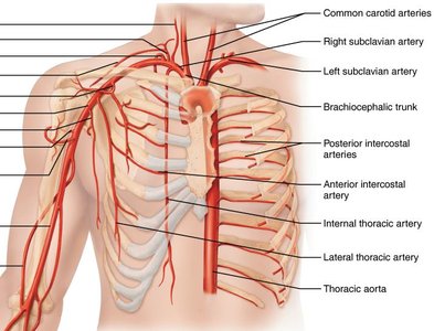
The aortic arch gives rise to several major arteries that supply the upper body.
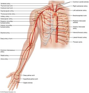
Brachiocephalic trunk: Only present on the right side; branches into right subclavian and right common carotid arteries.
Left subclavian artery: Direct branch from the aortic arch.
Common carotid arteries: Supply the head and neck.
Arteries of the Head and Neck
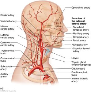
The head and neck are supplied by branches of the external and internal carotid arteries.
External carotid artery: Supplies superficial structures of the head and neck.
Internal carotid artery: Supplies the brain.
Arteries of the Upper Limb
The upper limb receives blood from the subclavian, axillary, brachial, radial, and ulnar arteries.
Axillary artery: Located in the armpit region.
Brachial artery: Main artery of the upper arm.
Radial and ulnar arteries: Supply the forearm and hand.
Abdominal Arteries and Celiac Trunk
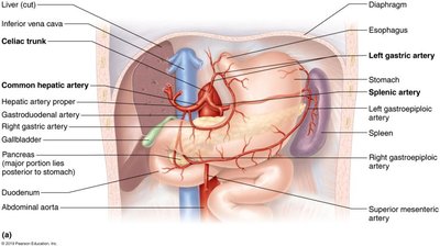
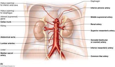
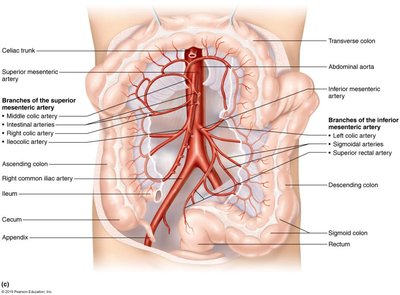
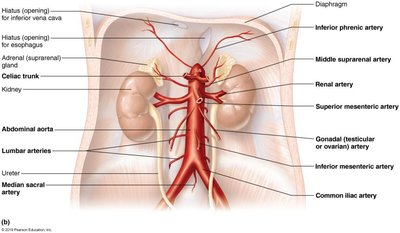
The abdominal aorta gives rise to several important branches, including the celiac trunk, which supplies the stomach, liver, spleen, and other organs.
Celiac trunk: Branches into left gastric, splenic, and common hepatic arteries.
Superior mesenteric artery: Supplies the small intestine and part of the large intestine.
Inferior mesenteric artery: Supplies the distal large intestine.
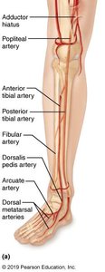
Hepatic Portal System
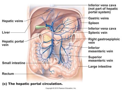
The hepatic portal system is a unique vascular circuit that carries blood from the gastrointestinal organs to the liver for filtration before it enters the systemic circulation.
Hepatic portal vein: Collects blood from the intestines, stomach, and spleen.
Hepatic veins: Carry filtered blood from the liver to the inferior vena cava.
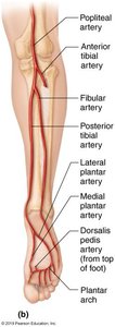
Blood Supply to the Foot
The foot receives blood from branches of the popliteal artery, including the anterior tibial, posterior tibial, and fibular arteries. The dorsalis pedis artery is a continuation of the anterior tibial artery.
Anterior tibial artery: Supplies the anterior compartment of the leg and dorsum of the foot.
Posterior tibial artery: Supplies the posterior compartment of the leg and plantar surface of the foot.
Fibular artery: Supplies the lateral compartment of the leg.
Fetal Circulation
Fetal circulation differs from adult circulation due to the presence of unique structures that bypass certain organs. These structures close after birth.
Ductus arteriosus: Connects the pulmonary artery to the aorta; closes to become ligamentum arteriosum.
Foramen ovale: Opening between the right and left atria; closes to become fossa ovalis.
Patent: Means the structure remains open when it should close.
Structure | Fetal State | Adult State |
|---|---|---|
Ductus arteriosus | Open | Ligamentum arteriosum (closed) |
Foramen ovale | Open | Fossa ovalis (closed) |
Summary Table: Major Arteries and Veins
Region | Major Arteries | Major Veins |
|---|---|---|
Head/Neck | Common carotid, internal carotid, external carotid | Jugular veins |
Thorax | Brachiocephalic trunk, subclavian, thoracic aorta | Brachiocephalic vein, superior vena cava |
Abdomen | Abdominal aorta, celiac trunk, mesenteric arteries | Inferior vena cava, hepatic veins, portal vein |
Upper Limb | Axillary, brachial, radial, ulnar | Cephalic, basilic, brachial |
Lower Limb | Femoral, popliteal, tibial, fibular | Femoral, popliteal, saphenous |
Key Terms and Concepts
Collateral circulation: Alternative pathways for blood flow in case of vessel occlusion.
Regional naming: Vessels are named according to the region they supply or drain.
Portal system: A vascular system where blood passes through two capillary beds before returning to the heart.
Example: Tracing Blood Flow
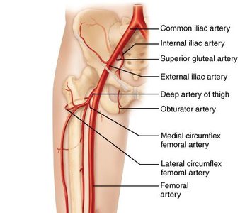
To trace blood flow from the heart to the foot: Aorta → Common iliac artery → External iliac artery → Femoral artery → Popliteal artery → Anterior/posterior tibial artery → Dorsalis pedis artery.
Additional info:
Some content was inferred from standard anatomical knowledge to ensure completeness and clarity.
