 뒤로
Bones and Bone Structure: Study Guide for Anatomy & Physiology
스터디 가이드 - 스마트 노트
 자료에 맞춘 맞춤형 노트, 핵심 정의, 예시, 맥락을 확장해 제공합니다.
Bones and Bone Structure
Functions of the Skeletal System
The skeletal system is essential for structural integrity and physiological function in the human body. It consists of bones, cartilages, ligaments, and other connective tissues that stabilize and interconnect bones.
Structural Support: Provides the framework for the body and supports soft tissues.
Storage of Minerals and Lipids: Stores calcium, phosphorus, and lipids in yellow bone marrow.
Blood Cell Production: Red bone marrow produces red and white blood cells and platelets.
Protection: Shields vital organs and soft tissues (e.g., skull protects the brain).
Leverage: Bones act as levers for muscles, enabling movement.
Classification of Bones
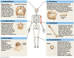
Bones are classified by their shape and structure, which relates to their function and location in the body.
Sutural Bones: Small, flat, irregular bones found between skull bones. Number varies among individuals.
Irregular Bones: Complex shapes (e.g., vertebrae, pelvic bones).
Short Bones: Boxy, found in wrists (carpals) and ankles (tarsals).
Flat Bones: Thin, parallel surfaces (e.g., skull roof, sternum, ribs, scapulae).
Long Bones: Long and slender, found in limbs (e.g., humerus, femur).
Sesamoid Bones: Small, round, flat bones within tendons (e.g., patella).
Bone Markings (Surface Features)
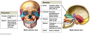
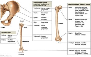
Bone markings are structural features that serve as sites for muscle attachment, articulation, or passage of nerves and blood vessels.
Projections: For attachment of muscles, tendons, ligaments, or articulation with other bones.
Openings and Depressions: For passage of blood vessels and nerves.
Structure of Long and Flat Bones
Long bones and flat bones have distinct structural features that support their function.
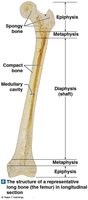
Long Bone:
Diaphysis: Tubular shaft made of compact bone surrounding a medullary cavity.
Epiphysis: Ends of the bone, mostly spongy bone.
Metaphysis: Narrow zone connecting diaphysis and epiphysis.
Flat Bone:
Core of spongy bone (diploë) between two layers of compact bone (cortex).
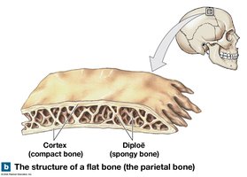
Bone Tissue (Osseous Tissue)
Bone tissue is a dense connective tissue composed of specialized cells and a solid extracellular matrix.
Matrix:
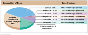
Two-thirds calcium phosphate (as hydroxyapatite crystals) – hard, brittle, resists compression.
One-third collagen fibers – strong, flexible, provides organic framework.
Cells:
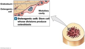
Osteogenic (osteoprogenitor) cells: Stem cells that differentiate into osteoblasts; important for fracture repair.
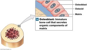
Osteoblasts: Immature cells responsible for osteogenesis (bone matrix production); become osteocytes when surrounded by matrix.
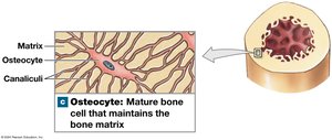
Osteocytes: Mature bone cells in lacunae; maintain matrix and help repair bone.
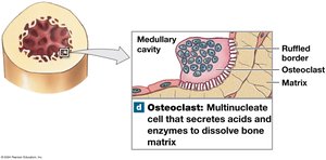
Osteoclasts: Large, multinucleate cells that absorb and remove bone matrix (osteolysis); secrete acids and enzymes.
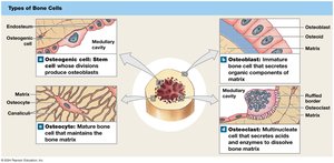
Compact Bone and Spongy Bone
Compact and spongy bone have distinct structures and functions.
Compact Bone:
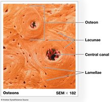
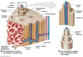
Basic unit: Osteon (central canal with blood vessels, concentric lamellae, osteocytes in lacunae).
Types of lamellae: concentric (osteons), interstitial (between osteons), circumferential (outer/inner surfaces).
Spongy Bone:
No osteons; matrix arranged as trabeculae.
Contains red bone marrow (blood cell production) and sometimes yellow bone marrow (fat storage).
Supports and protects marrow, withstands stress from many directions.
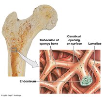
Coordinated Functions of Compact and Spongy Bone
Compact and spongy bone work together to bear weight and resist forces.
Trabeculae in spongy bone transfer weight to compact bone.
Medial shaft compresses; lateral shaft resists bending (tension).
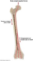
Surface Coverings of Bone
Bones are covered by specialized membranes that support growth, repair, and attachment.
Periosteum: Outer fibrous and inner cellular layers; continuous with joint capsule and tendons; isolates bone, provides route for vessels/nerves, participates in growth/repair.
Perforating fibers (Sharpey’s fibers): Collagen fibers that increase strength of attachment.
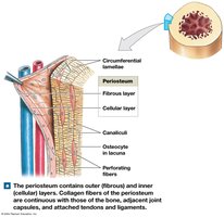
Endosteum: Incomplete cellular layer lining inner surfaces; contains osteogenic cells, osteoblasts, osteoclasts; active during growth, repair, remodeling.
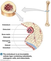
Bone Formation and Growth
Bone formation (ossification) occurs via two mechanisms: endochondral and intramembranous ossification.
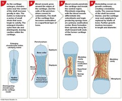
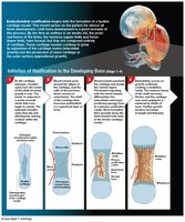
Endochondral Ossification: Bone replaces cartilage. Steps include:
Chondrocytes enlarge, matrix calcifies, chondrocytes die.
Blood vessels grow around cartilage, osteoblasts form bone collar.
Blood vessels enter, osteoblasts produce spongy bone.
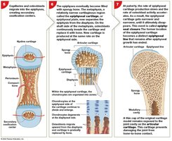
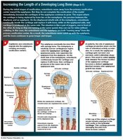
Spongy bone remodels, marrow cavity forms.
Secondary ossification centers form in epiphyses.
Epiphyses fill with spongy bone, epiphyseal plate separates diaphysis/epiphysis.
Epiphyseal closure at puberty; epiphyseal line remains.
Bone Growth:
Interstitial growth (length): New cartilage grows at epiphyseal plate, replaced by bone.
Appositional growth (width): Osteoblasts add lamellae, osteoclasts enlarge medullary cavity.
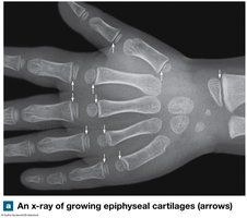
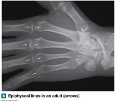
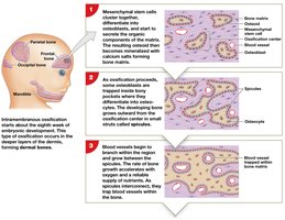
Intramembranous Ossification: Bone forms from mesenchymal cells in deeper dermis. Steps include:
Mesenchymal cells differentiate into osteoblasts, secrete osteoid.
Osteoblasts become osteocytes, bone grows in spicules.
Blood vessels grow between spicules, bone growth accelerates.
Osteoblasts deposit bone, plate of spongy bone forms.
Remodeling produces compact bone, periosteum forms.
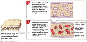
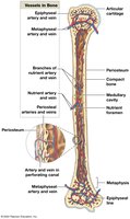
Blood Supply to Bones
Bones have an extensive blood supply, essential for growth, repair, and maintenance.
Nutrient artery and vein: Supply diaphysis.
Metaphyseal vessels: Supply epiphyseal cartilages.
Periosteal vessels: Supply superficial osteons and secondary ossification centers.
Lymphatic vessels: Enter bone via periosteum.
Sensory nerves: Innervate periosteum, endosteum, medullary cavity, epiphyses.
Bone Remodeling
Bone remodeling is a continuous process involving osteocytes, osteoblasts, and osteoclasts. It maintains bone integrity and adapts to stress.
Balanced activity keeps bones healthy.
Excess osteoclast activity weakens bones; excess osteoblast activity strengthens bones.
Exercise, Nutrition, and Hormones
Bone development and maintenance depend on physical activity, nutrients, and hormones.
Exercise: Weight-bearing activity increases bone strength; inactivity causes bone loss.
Minerals: Calcium, phosphorus, magnesium, fluoride, iron, manganese are essential.
Vitamins: D (calcitriol), C, A, K stimulate bone growth and maintenance.
Hormones: Growth hormone, thyroxine, sex hormones, parathyroid hormone, calcitonin regulate bone growth and calcium homeostasis.
Abnormal Bone Development
Pituitary growth failure: Short bones due to low growth hormone.
Gigantism: Tall stature from excess growth hormone before puberty.
Acromegaly: Thick bones from excess growth hormone after epiphyseal closure.
Marfan syndrome: Excess cartilage formation, tall stature, long limbs.
Calcium Homeostasis
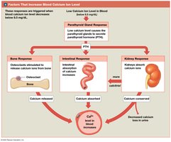
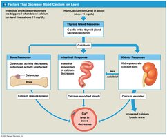
Bones store 99% of the body's calcium, which is vital for physiological processes. Calcium levels are regulated by hormones.
Parathyroid hormone (PTH): Increases blood calcium by stimulating osteoclasts, increasing intestinal absorption, decreasing kidney excretion.
Calcitonin: Decreases blood calcium by inhibiting osteoclasts, increasing kidney excretion, decreasing intestinal absorption.
Osteomalacia: Weak, flexible bones from poor mineralization.
Rickets: Osteomalacia due to vitamin D deficiency.
Fractures and Repair
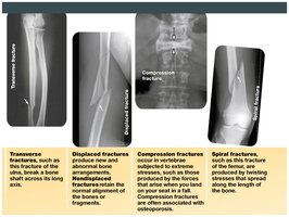
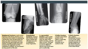
Fractures are cracks or breaks in bones due to physical stress. They are classified by their characteristics and location.
Open (compound) fracture: Projects through skin.
Closed (simple) fracture: Internal.
Other types: Transverse, displaced, compression, spiral, epiphyseal, comminuted, greenstick, Colles, Pott’s fracture.
Steps in Fracture Repair
Fracture hematoma formation: Blood clot closes vessels, bone cells die.
Callus formation: Endosteum and periosteum cells form internal (spongy bone) and external (cartilage and bone) calluses.
Spongy bone formation: Osteoblasts replace cartilage with spongy bone.
Compact bone formation: Calluses remodel and disappear; repaired bone may be thicker and stronger.
Effects of Aging on Bones
Aging leads to reduced bone mass and increased risk of fractures.
Osteopenia: Inadequate ossification, begins between ages 30-40; women lose more bone mass than men.
Osteoporosis: Severe bone loss, brittle bones, common in postmenopausal women; can be secondary to cancer.
Bone Cell Type | Function |
|---|---|
Osteogenic cells | Stem cells for osteoblasts; fracture repair |
Osteoblasts | Produce new bone matrix (osteogenesis) |
Osteocytes | Maintain matrix; repair bone |
Osteoclasts | Absorb/remove bone matrix (osteolysis) |
Hormone | Effect on Calcium |
|---|---|
Parathyroid hormone (PTH) | Increases blood calcium |
Calcitonin | Decreases blood calcium |
Bone Type | Example | Location |
|---|---|---|
Sutural | Wormian bones | Skull |
Irregular | Vertebrae | Spine |
Short | Carpals | Wrist |
Flat | Sternum | Chest |
Long | Femur | Thigh |
Sesamoid | Patella | Knee |
Additional info: Tables and images were recreated and expanded for clarity and completeness. All equations and formulas are provided in LaTeX format as required.
