뒤로
Bones and Bone Structure: Study Notes for Anatomy & Physiology
스터디 가이드 - 스마트 노트
 자료에 맞춘 맞춤형 노트, 핵심 정의, 예시, 맥락을 확장해 제공합니다.
Bones and Bone Structure
Functions of the Skeletal System
The skeletal system is essential for the structure, protection, and function of the human body. It consists of bones, cartilages, ligaments, and other connective tissues.
Support: Provides a framework for the body and supports soft tissues.
Storage of Minerals and Lipids: Bones store calcium and phosphate ions, as well as lipids in yellow marrow.
Blood Cell Production: Red marrow produces red blood cells, white blood cells, and platelets.
Protection: Bones protect vital organs (e.g., skull protects the brain, ribs protect the heart and lungs).
Leverage: Bones act as levers for muscles, enabling movement.
Classification of Bones
Bones are classified by their shape and structure, which relate to their function and location in the body.
Sutural Bones: Small, flat, irregular bones found between the flat bones of the skull.
Irregular Bones: Complex shapes (e.g., vertebrae, pelvic bones).
Short Bones: Boxlike in appearance (e.g., carpal and tarsal bones).
Flat Bones: Thin, parallel surfaces (e.g., skull, sternum, ribs, scapulae).
Long Bones: Long and slender (e.g., femur, humerus, fingers, toes).
Sesamoid Bones: Small, round, and flat; found near joints (e.g., patella).
Structure of a Long Bone
Long bones have a characteristic structure that supports their function in movement and weight-bearing.
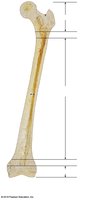
Diaphysis: The shaft; composed of compact bone surrounding the medullary (marrow) cavity.
Epiphysis: The expanded ends; mostly spongy bone covered by compact bone.
Metaphysis: The region where the diaphysis and epiphysis meet; site of growth during development.
Blood Supply to Bone
Bones are highly vascularized, receiving blood through several types of vessels:
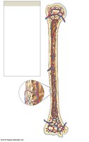
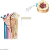
Nutrient Artery and Vein: Enter through the nutrient foramen to supply the diaphysis and medullary cavity.
Metaphyseal and Epiphyseal Vessels: Supply the metaphysis and epiphysis, respectively.
Periosteal Vessels: Supply the superficial osteons and periosteum.
Compact Bone and Spongy Bone
Bones consist of two main types of osseous tissue: compact bone and spongy bone.
Compact Bone
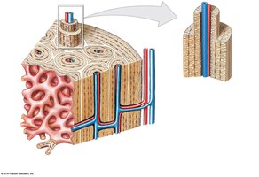
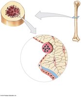
Osteon: The functional unit; consists of concentric lamellae around a central canal containing blood vessels.
Perforating Canals: Carry blood vessels perpendicular to the bone surface.
Lamellae: Layers of bone matrix; can be concentric, interstitial, or circumferential.
Spongy Bone
Trabeculae: Network of struts and plates; lacks osteons.
Red Bone Marrow: Fills spaces between trabeculae; site of blood cell formation.
Yellow Bone Marrow: Stores fat; found in other sites of spongy bone.
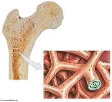
Distribution of Forces on a Long Bone
Long bones are adapted to withstand forces. The trabeculae in the epiphysis transfer forces from the pelvis to the shaft, with compression on the medial side and tension on the lateral side.
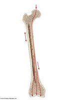
Periosteum and Endosteum
Bone surfaces are covered by specialized membranes:
Periosteum: Covers the outer surface (except at joints); has an outer fibrous layer and inner cellular layer; provides attachment for tendons and ligaments.
Endosteum: Lines the medullary cavity and covers trabeculae; contains osteogenic cells, osteoblasts, and osteoclasts; active in bone growth and repair.
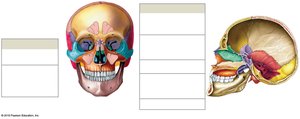
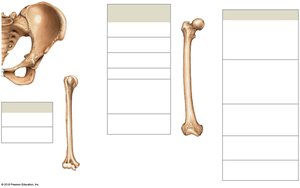
Bone Markings (Surface Features)
Bones have characteristic markings that serve as sites for muscle attachment, articulation, and passage of nerves and vessels.
Type | Example/Function |
|---|---|
Projections | Process, ramus, trochanter, crest, spine, line, tubercle, tuberosity, head, neck, facet, condyle, trochlea |
Openings | Foramen, canal, meatus, fissure, sinus |
Depressions | Fossa, sulcus |
Bone (Osseous) Tissue and Bone Cells
Bone tissue is a dense, supportive connective tissue with a solid matrix and specialized cells.
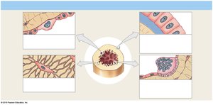
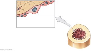
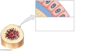
Osteogenic Cells: Stem cells that divide to produce osteoblasts; found in periosteum and endosteum; important for fracture repair.
Osteoblasts: Immature cells that produce new bone matrix (osteoid); become osteocytes when surrounded by matrix.
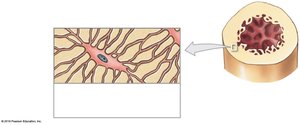
Osteocytes: Mature bone cells; maintain bone matrix and help repair damaged bone; reside in lacunae and communicate via canaliculi.
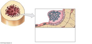
Osteoclasts: Large, multinucleate cells that dissolve bone matrix (osteolysis); important for calcium homeostasis.
Bone Formation and Growth
Bone development involves two main processes: ossification and calcification.
Ossification (Osteogenesis): Formation of bone tissue.
Calcification: Deposition of calcium salts during ossification.
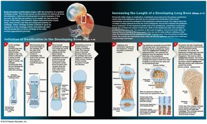
Endochondral Ossification: Most bones form this way; bone replaces hyaline cartilage in seven main steps.
Intramembranous Ossification: Bone develops directly from mesenchymal tissue; forms dermal bones (e.g., mandible, clavicle).
Bone Growth
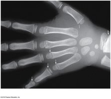
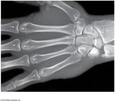
Interstitial Growth: Growth in length; occurs at epiphyseal plates until closure (epiphyseal line remains).
Appositional Growth: Growth in width; new layers of bone are added to the surface, and the medullary cavity enlarges.
Calcium Homeostasis
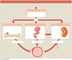
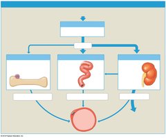
Bones store 99% of the body's calcium, which is vital for many physiological processes. Calcium ion concentrations are regulated by hormones:
Parathyroid Hormone (PTH): Increases blood calcium by stimulating osteoclasts, increasing intestinal absorption (via calcitriol), and reducing kidney excretion.
Calcitonin: Decreases blood calcium by inhibiting osteoclasts, increasing kidney excretion, and reducing intestinal absorption.
Bone Remodeling
Bone remodeling is a continuous process involving osteocytes, osteoblasts, and osteoclasts. It maintains bone strength and mineral homeostasis.
Balanced Activity: Bone deposition and resorption are normally balanced.
Imbalance: Excessive resorption weakens bones; excessive deposition strengthens bones.
Exercise, Nutrition, and Hormones
Bone health depends on physical activity, nutrition, and hormonal regulation.
Exercise: Weight-bearing exercise stimulates osteoblasts and increases bone density.
Nutrition: Adequate intake of calcium, phosphorus, magnesium, fluoride, iron, manganese, vitamins D3, C, A, K, and B12 is essential.
Hormones: Growth hormone, thyroxine, estrogen, testosterone, PTH, and calcitonin all influence bone growth and maintenance.
Effects of Aging on the Skeletal System
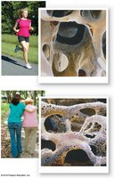
Aging leads to decreased bone mass and increased risk of fractures.
Osteopenia: Inadequate ossification; begins between ages 30 and 40.
Osteoporosis: Severe bone loss; more common in women, especially after menopause.
Consequences: Fragile limbs, reduced height, tooth loss.
Fractures and Bone Repair
Fractures are breaks in bone due to physical stress. They are classified as open (compound) or closed (simple), with several specific types (e.g., transverse, comminuted, greenstick).
Steps in Fracture Repair
Fracture Hematoma Formation: Blood clot forms at the site; bone cells die.
Callus Formation: Internal and external calluses form to stabilize the break.
Spongy Bone Formation: Osteoblasts replace cartilage with spongy bone.
Compact Bone Formation: Spongy bone is replaced by compact bone; bone may be slightly thicker at the repair site.