 뒤로
뒤로Bones and Bone Tissue: Structure, Classification, and the Axial Skeleton
스터디 가이드 - 스마트 노트
 자료에 맞춘 맞춤형 노트, 핵심 정의, 예시, 맥락을 확장해 제공합니다.
Bones and Cartilage: Overview and Classification
Axial vs. Appendicular Skeleton
The human skeleton is divided into two main parts: the axial skeleton and the appendicular skeleton. The axial skeleton forms the central axis of the body and includes the skull, vertebral column, and thoracic cage. The appendicular skeleton consists of the bones of the limbs and girdles that attach them to the axial skeleton.
Axial skeleton: Skull, vertebral column, ribs, sternum, and hyoid bone.
Appendicular skeleton: Pectoral girdle, pelvic girdle, upper and lower limb bones.
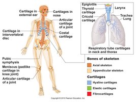
Types of Cartilage in the Skeleton
Cartilage is a resilient and smooth elastic tissue that covers and protects the ends of long bones at the joints and is a structural component of the rib cage, ear, nose, bronchial tubes, intervertebral discs, and many other body components.
Hyaline cartilage: Most abundant; found in articular surfaces, costal cartilage, nose, trachea, and larynx.
Elastic cartilage: Found in the external ear and epiglottis.
Fibrocartilage: Found in intervertebral discs, pubic symphysis, and menisci of the knee.
Classification of Bones by Shape
Bone Shapes
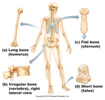
Bones are classified according to their shapes, which relate to their functions.
Long bones: Longer than they are wide (e.g., humerus, femur).
Short bones: Nearly equal in length and width (e.g., carpals, tarsals).
Flat bones: Thin, flattened, and usually curved (e.g., sternum, scapulae, ribs, skull bones).
Irregular bones: Complicated shapes (e.g., vertebrae, some facial bones).
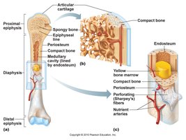
Structure of a Long Bone
Gross Anatomy of Long Bones
Long bones have a complex structure that supports their function in movement and support.
Diaphysis: The shaft of the bone, composed mainly of compact bone.
Epiphyses: The ends of the bone, consisting mostly of spongy bone covered by compact bone.
Medullary cavity: Central cavity within the diaphysis, containing yellow bone marrow in adults.
Epiphyseal line: Remnant of the epiphyseal plate, a region of cartilage where bone growth occurs in children.
Articular cartilage: Hyaline cartilage covering the joint surfaces of epiphyses.
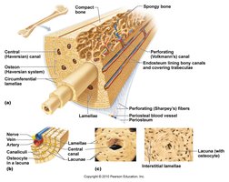
Microscopic Structure of Bone Tissue
Compact Bone
Compact bone is dense and forms the outer layer of all bones, providing strength for weight bearing.
Osteon (Haversian system): The structural unit of compact bone, consisting of concentric lamellae around a central canal.
Lamellae: Layers of bone matrix.
Central (Haversian) canal: Contains blood vessels and nerves.
Lacunae: Small spaces housing osteocytes.
Canaliculi: Tiny canals connecting lacunae, allowing for nutrient and waste exchange.
Perforating (Volkmann's) canals: Connect blood vessels and nerves of the periosteum to those in the central canals.
Bone Markings and Features
Terms for Bone Features
Bones have various surface features that serve as sites for muscle, ligament, and tendon attachment, or as passages for nerves and blood vessels.
Feature | Description |
|---|---|
Process | Any bony prominence |
Foramen | Round or oval opening through a bone |
Fossa | Shallow, basinlike depression in a bone |
Condyle | Rounded articular projection |
Crest | Narrow, prominent ridge of bone |
Spine | Sharp, slender, often pointed projection |
Tubercle/Tuberosity | Small/large rounded projection |
Meatus | Canal-like passageway |
Sinus | Cavity within a bone, filled with air and lined with mucous membrane |
Head | Bony expansion carried on a narrow neck |
*Additional info: See Table 7.3 in your textbook for a comprehensive list of skull features and their descriptions.* 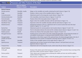
Bones of the Skull: Orbit and Nasal Cavity
Bones Forming the Orbit
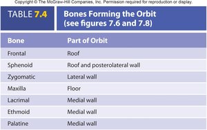
The orbit is a bony cavity that houses the eyeball and associated structures. It is formed by seven bones.
Bone | Part of Orbit |
|---|---|
Frontal | Roof |
Sphenoid | Roof and posterolateral wall |
Zygomatic | Lateral wall |
Maxilla | Floor |
Lacrimal | Medial wall |
Ethmoid | Medial wall |
Palatine | Medial wall |
Bones Forming the Nasal Cavity
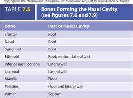
The nasal cavity is formed by several bones that create the roof, floor, septum, and lateral walls.
Bone | Part of Nasal Cavity |
|---|---|
Frontal | Roof |
Nasal | Roof |
Sphenoid | Roof |
Ethmoid | Roof, septum, lateral wall |
Inferior nasal concha | Lateral wall |
Lacrimal | Lateral wall |
Maxilla | Floor |
Palatine | Floor and lateral wall |
Vomer | Septum |
Paranasal Sinuses
Functions and Locations
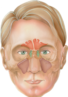
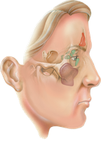
Paranasal sinuses are air-filled spaces within certain skull bones. They serve to lighten the skull, act as resonating chambers for voice, and produce mucus.
Frontal sinus
Maxillary sinus
Ethmoidal sinus
Sphenoidal sinus
Sutures of the Skull
Major Sutures
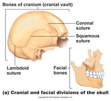
Sutures are immovable joints found only between skull bones. They fuse the bones of the cranium and face.
Coronal suture: Between frontal and parietal bones
Sagittal suture: Between right and left parietal bones
Lambdoid suture: Between parietal and occipital bones
Squamous suture: Between parietal and temporal bones
Inferior and Internal Views of the Skull
Inferior View
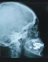
The inferior view of the skull reveals the base, including the hard palate, foramen magnum, and various foramina for nerves and blood vessels.
Internal View (Cranial Fossae)
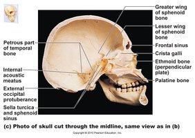
The cranial cavity is divided into three fossae: anterior, middle, and posterior, which accommodate different parts of the brain.
Anterior cranial fossa: Supports the frontal lobes of the brain
Middle cranial fossa: Supports the temporal lobes
Posterior cranial fossa: Supports the cerebellum
Summary Table: Processes and Features of the Skull
The following table summarizes key processes and features of the skull, their locations, and descriptions.
Summary Table: Bones Forming the Orbit
Summary Table: Bones Forming the Nasal Cavity
