 뒤로
Cardiac Function and Electrical Activity: Anatomy & Physiology Study Guide
스터디 가이드 - 스마트 노트
 자료에 맞춘 맞춤형 노트, 핵심 정의, 예시, 맥락을 확장해 제공합니다.
The Cardiovascular System: Cardiac Function
System Overview
The cardiovascular system (CVS) is essential for transporting blood throughout the body, ensuring the delivery of oxygen, nutrients, and removal of wastes. It consists of three principal components:
Heart: The muscular pump that propels blood.
Blood vessels: The network of pipes (arteries, veins, capillaries) through which blood flows.
Blood: The fluid medium carrying gases, nutrients, and wastes.
CVS function is regulated by the endocrine system, nervous system, and kidneys.
Heart Structure and Circuits
The heart is a four-chambered organ, acting as a double pump for two main circuits:
Pulmonary circuit: Carries O2-deficient blood from the heart to the lungs and back.
Systemic circuit: Delivers O2-rich blood from the heart to the rest of the body and returns deoxygenated blood.
Coronary circuit: Supplies the heart muscle (myocardium) with blood.
The heart wall consists of three layers:
Epicardium: Outer layer (visceral and parietal pericardium).
Myocardium: Middle layer of cardiac muscle tissue.
Endocardium: Inner layer, continuous with blood vessel endothelium.
Heart Anatomy
The heart's chambers and major vessels are organized to facilitate efficient blood flow. The right ventricle pumps blood to the lungs (pulmonary circuit), while the left ventricle pumps blood to the systemic circuit. The left ventricle has a thicker wall to generate higher pressure for systemic circulation.
Blood Flow Pathways
Blood flows in series through the heart and circuits:
Left ventricle → aorta → systemic circuit → vena cavae → right atrium → right ventricle → pulmonary artery → pulmonary circuit → pulmonary veins → left atrium → left ventricle
Equal volumes of blood are pumped to both circuits, but the systemic circuit is longer and higher pressure.
Coronary Circulation
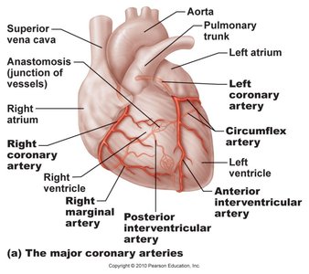
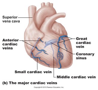
The heart muscle receives its own blood supply via the coronary arteries aនd returns deoxygenated blood through cardiac veins. Coronary circulation is vital for myocardial function.
Arteries | Veins |
|---|---|
Right & left coronary, marginal, circumflex, anterior interventricular | Small cardiac, anterior cardiac, great cardiac veins |
Heart Valves
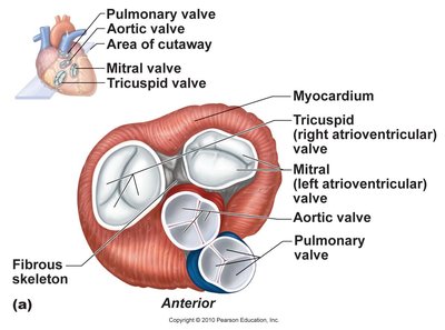
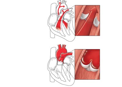
Valves ensure unidirectional blood flow:
Atrioventricular (AV) valves: Tricuspid (right), Mitral (left); prevent backflow into atria.
Semilunar (SL) valves: Aortic and pulmonary; prevent backflow into ventricles.
Valve Function During Cardiac Cycle
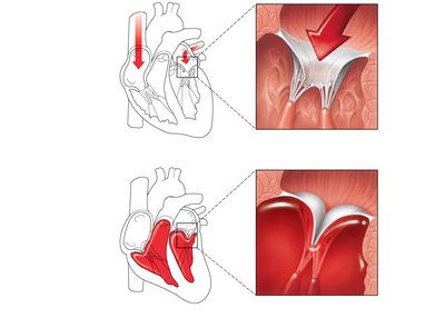
AV valves open when atrial pressure exceeds ventricular pressure, allowing blood to flow into ventricles. They close during ventricular contraction to prevent backflow. SL valves open during ventricular contraction and close during relaxation.
Heart Sounds and Clinical Assessment
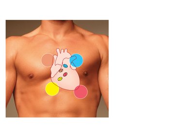
Heart sounds ("lub-dup") are associated with valve closure:
First sound: AV valves close (beginning of systole).
Second sound: SL valves close (beginning of diastole).
Heart murmurs indicate abnormal valve function. Auscultation points for valve sounds are clinically important.
Cardiac Muscle and Conduction System
Cardiac Muscle Tissue
Cardiac muscle cells (cardiocytes) differ from skeletal muscle:
Depend on aerobic respiration.
Short T-tubules, extensive blood supply.
Non-voluntary contraction.
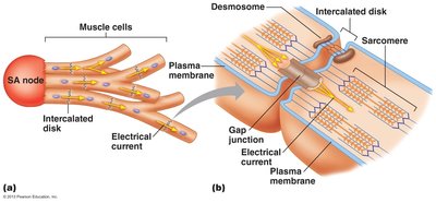
Interconnected by intercalated discs (gap junctions and desmosomes).
The Conduction System of the Heart
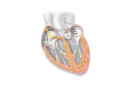
The heart's electrical activity is coordinated by specialized autorhythmic cells:
Sinoatrial (SA) node: Pacemaker, initiates impulses.
Atrioventricular (AV) node: Delays impulse, allows ventricular filling.
AV bundle (bundle of His): Connects atria and ventricles.
Bundle branches: Conduct impulses through interventricular septum.
Purkinje fibers: Depolarize ventricular muscle.
Location | Firing Rate at Rest (AP/min) |
|---|---|
SA node | 70–80 |
AV node | 40–60 |
Bundle of His | 20–40 |
Purkinje fibers | 20–40 |
Ionic Basis of Electrical Activity
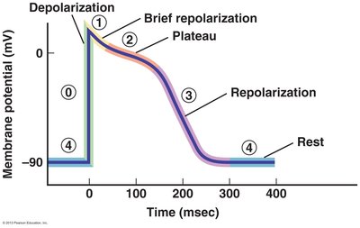
Cardiac contractile cells undergo five phases of electrical activity:
Phase 0: Increased Na+ permeability (depolarization).
Phase 1: Decreased Na+ permeability (brief repolarization).
Phase 2: Increased Ca2+ permeability, decreased K+ permeability (plateau).
Phase 3: Increased K+ permeability, decreased Ca2+ permeability (repolarization).
Phase 4: Resting membrane potential.
Recording the Electrical Activity of the Heart
Electrocardiogram (ECG or EKG)
An ECG records the electrical changes during the cardiac cycle. It is a composite of all action potentials generated by nodal and contractile cells. ECGs are used to detect conduction abnormalities, myocardial infarction, and arrhythmias.
P wave: Depolarization of SA node (atrial excitation).
QRS complex: Ventricular depolarization (ventricular excitation).
T wave: Ventricular repolarization (ventricular relaxation).
ECG Interpretation
Clinicians analyze:
P wave and QRS complex size & duration
ST segment
Intervals (P-Q, S-T, Q-T)
ECG abnormalities include:
Sinus rhythm: Normal pace by SA node.
Tachycardia: Fast rhythm.
Bradycardia: Slow rhythm.
Heart blocks: Impaired conduction at AV node (1st, 2nd, 3rd degree).
Extra contractions: Premature atrial or ventricular contractions.
Ventricular fibrillation: Loss of coordination, potentially fatal.
Summary Table: Cardiac Conduction and ECG Waves
Wave | Event | Clinical Significance |
|---|---|---|
P wave | Atrial depolarization | SA node function |
QRS complex | Ventricular depolarization | Ventricular function |
T wave | Ventricular repolarization | Recovery phase |
Key Equations
Cardiac Output (CO):
Blood Flow:
Where is the pressure difference and is resistance.
Clinical Connections
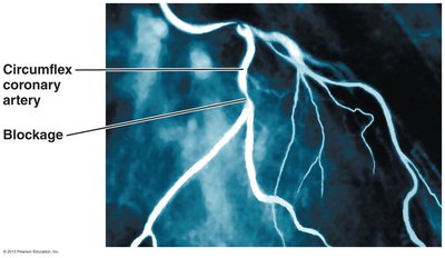
Coronary Artery Blockage
Blockages in coronary arteries can lead to myocardial infarction (heart attack). Angiograms are used to visualize these blockages.
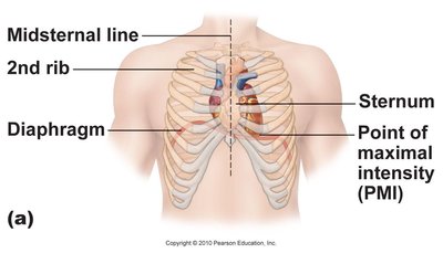
Heart Sounds and Auscultation
Proper placement of stethoscope is essential for detecting heart sounds and murmurs.
Conclusion
The cardiac function and electrical activity are central to understanding the physiology of the heart. Mastery of these concepts is essential for clinical assessment and diagnosis of cardiovascular diseases.
