 뒤로
Cardiac Function and Electrical Activity: Anatomy & Physiology Study Notes
스터디 가이드 - 스마트 노트
 자료에 맞춘 맞춤형 노트, 핵심 정의, 예시, 맥락을 확장해 제공합니다.
The Cardiovascular System: Cardiac Function
System Overview
The cardiovascular system (CVS) is essential for transporting blood throughout the body, delivering oxygen, nutrients, and removing waste products. It consists of three principal components:
Heart: The muscular pump that propels blood.
Blood Vessels: The network of pipes (arteries, veins, capillaries) through which blood flows.
Blood: The fluid medium carrying cells and dissolved substances.
CVS function is influenced by the endocrine system, nervous system, and kidneys, which regulate blood pressure, volume, and composition.
Heart Structure and Circuits
The heart is a small organ, roughly the size of a clenched fist, with four muscular chambers: right and left atria, and right and left ventricles. It acts as a double pump, supporting three circuits:
Pulmonary Circuit: Carries O2-deficient blood from the heart to the lungs and back.
Systemic Circuit: Delivers O2-rich blood from the heart to the rest of the body and returns it.
Coronary Circuit: Supplies the heart muscle (myocardium) with blood.
The heart wall consists of three layers:
Epicardium: Outer layer (visceral and parietal pericardium).
Myocardium: Middle layer of cardiac muscle tissue, blood vessels, and nerves.
Endocardium: Inner layer (simple squamous epithelium), continuous with blood vessel endothelium.
Heart Anatomy
The heart's anatomy includes the four chambers, valves, and major blood vessels. The right ventricle pumps blood to the lungs (pulmonary circuit) with low pressure, while the left ventricle pumps blood to the systemic circuit with higher pressure due to greater resistance and distance.
Right Ventricle: Thin wall, low pressure, short distance.
Left Ventricle: Thick wall, high pressure, long distance.
Equal volumes of blood are pumped to both circuits, but the anatomy of the ventricles reflects their functional differences.
Path of Blood Flow
The cardiovascular system is a closed system, with blood flowing in series through the pulmonary and systemic circuits:
Left ventricle → aorta → systemic circuit → vena cavae → right atrium → right ventricle → pulmonary artery → pulmonary circuit → pulmonary veins → left atrium → left ventricle
In pulmonary capillary beds, O2 moves into the blood and CO2 leaves. In systemic capillary beds, O2 leaves the blood and CO2 enters.
Coronary Circulation
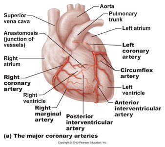
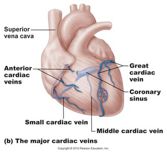
The coronary circuit provides the functional blood supply to the heart muscle itself. It includes:
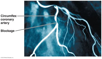
Arteries: Right and left coronary, marginal, circumflex, anterior interventricular arteries.
Veins: Small cardiac, anterior cardiac, great cardiac veins.
Collateral routes and anastomoses ensure additional blood delivery pathways.
Heart Valves
Structure and Function
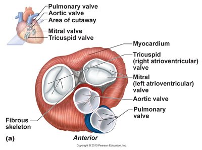
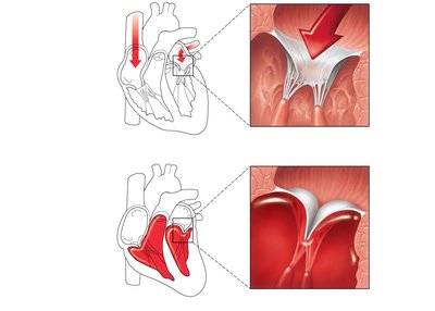
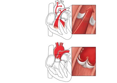
Heart valves ensure unidirectional blood flow through the heart. There are two main types:
Atrioventricular (AV) Valves: Prevent backflow into the atria when ventricles contract. Includes the tricuspid (right) and mitral (left) valves. Chordae tendineae anchor AV valve cusps to papillary muscles.
Semilunar (SL) Valves: Prevent backflow into the ventricles when ventricles relax. Includes the aortic and pulmonary semilunar valves.
Heart Sounds and Clinical Relevance
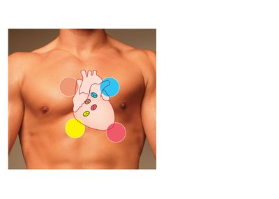
Two sounds (lub-dup) are associated with the closing of heart valves:
First sound: AV valves close, beginning of systole.
Second sound: SL valves close, beginning of ventricular diastole.
Heart murmurs are abnormal sounds, often indicative of valve problems.
Cardiac Muscle Tissue
Structure and Properties
Cardiac muscle cells (cardiocytes) differ from skeletal muscle fibers:
Depend almost entirely on aerobic respiration.
Short T-tubules, do not form triads with sarcoplasmic reticulum.
Extensive circulatory supply.
Non-voluntary contraction.
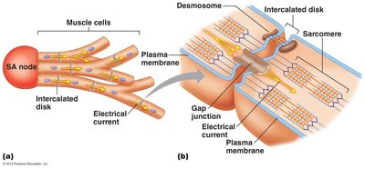
Interconnected by intercalated discs (specialized cell junctions).
The Conduction System of the Heart
Spread of Excitation
The heart's conduction system coordinates the spread of electrical excitation, ensuring the atria contract before the ventricles. This is achieved through gap junctions and conduction pathways:
Intercalated disks: Junctions between myocardial cells, containing desmosomes (mechanical stress resistance) and gap junctions (electrical coupling).
Autorhythmicity: The heart's ability to generate its own rhythm via autorhythmic cells.
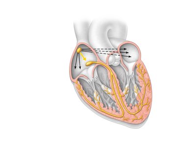
Intrinsic Cardiac Conduction System
A network of noncontractile (autorhythmic) cells initiates and distributes impulses to coordinate depolarization and contraction:
Sinoatrial (SA) node: Pacemaker, generates impulses (~75/min).
Atrioventricular (AV) node: Delays impulses (~0.1 s), depolarizes 50/min.
AV bundle (bundle of His): Only electrical connection between atria and ventricles.
Right and left bundle branches: Carry impulses toward the apex.
Purkinje fibers: Complete pathway into apex and ventricular walls.
Depolarization is rhythmic and spontaneous. Fastest depolarizing cells (SA node) set the rate for the rest of the heart.
Ionic Basis of Electrical Activity
Cardiac contractile cells undergo five phases of electrical activity:
Phase 0: Increased Na+ permeability (depolarization).
Phase 1: Decreased Na+ permeability (brief repolarization).
Phase 2: Increased Ca2+ permeability, decreased K+ permeability (plateau).
Phase 3: Increased K+ permeability, decreased Ca2+ permeability (repolarization).
Phase 4: Resting membrane potential.
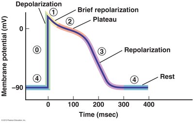
Equation: Resting membrane potential is typically in cardiac cells.
Recording the Electrical Activity of the Heart: Electrocardiogram (ECG)
Principles and Clinical Use
An ECG records the electrical changes during the cardiac cycle using electrodes on the skin. It is a noninvasive technique used to detect clinical abnormalities related to conduction, such as myocardial infarction (MI) or arrhythmias.
P wave: Depolarization of SA node (atrial excitation).
QRS complex: Ventricular depolarization (ventricular excitation).
T wave: Ventricular repolarization (ventricular relaxation).
Clinicians analyze the size and duration of the P wave and QRS complex, ST segment, and intervals to assess cardiac function, blood flow, hypertrophy, atrophy, or damage.
ECG Arrhythmias and Heart Blocks
Common arrhythmias include:
Sinus rhythm: Normal pace generated by SA node.
Tachycardia: Fast rhythm.
Bradycardia: Slow rhythm.
Heart blocks: Impaired conduction through AV node (1st, 2nd, 3rd degree).
Extra contractions: Premature atrial contraction (PAC), premature ventricular contraction (PVC).
Ventricular fibrillation: Loss of coordination of electrical activity, potentially fatal.
ECGs are divided into segments and intervals, representing voltage and time.
Summary Table: Cardiac Conduction System Firing Rates
Location | Firing Rate at Rest (AP/min) |
|---|---|
SA node | 70–80 |
AV node | 40–60 |
Bundle of His | 20–40 |
Purkinje fibers | 20–40 |
Example: An ECG can reveal a myocardial infarction by showing abnormal ST segment elevation.
Additional info: The notes have been expanded with academic context to ensure completeness and clarity for exam preparation.
