 뒤로
Cardiac Muscle: Structure, Function, and Physiology
스터디 가이드 - 스마트 노트
 자료에 맞춘 맞춤형 노트, 핵심 정의, 예시, 맥락을 확장해 제공합니다.
Cardiac Muscle
Structure and Cellular Organization
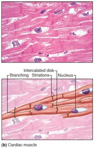
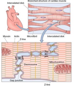
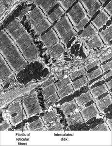
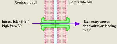
Cardiac muscle is a specialized form of muscle tissue found only in the heart. It is composed of cardiac myocytes, which are shorter, branched cells typically containing a single nucleus. These cells are interconnected by intercalated disks, which facilitate both mechanical and electrical coupling.
Cardiac myocytes: Short, branched, usually single-nucleated cells.
Intercalated disks: Specialized junctions containing desmosomes (mechanical linkage) and gap junctions (electrical linkage).
Striations: Cardiac muscle is striated, with sarcomeres containing thick (myosin) and thin (actin) filaments.
Mitochondria: Abundant, occupying about one-third of cell volume, supporting oxidative metabolism.
T-tubules: Larger and less abundant than in skeletal muscle.
Sarcoplasmic reticulum: Smaller, requiring extracellular Ca2+ entry for contraction.
Autorhythmic and Contractile Cells
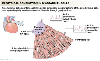
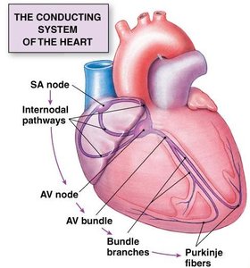
The heart contains two main types of myocardial cells: contractile cells (responsible for contraction) and autorhythmic cells (pacemaker cells, ~1% of total). Autorhythmic cells generate action potentials spontaneously, initiating the heartbeat.
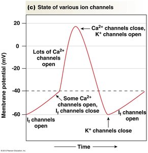
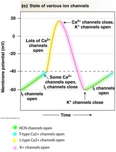
Autorhythmic cells: Located primarily in the sinoatrial (SA) node, have unstable resting membrane potentials (~-60 mV).
Contractile cells: Make up the majority of cardiac muscle, responsible for force generation.
HCN channels: Hyperpolarization-activated cyclic nucleotide-gated channels ("funny current") open at -60 mV, allowing Na+ influx and gradual depolarization.
T-type Ca2+ channels: Open just before threshold, further depolarizing the cell.
L-type Ca2+ channels: Open at threshold, causing the action potential spike.
K+ channels: Open to repolarize the cell.
Action Potentials in Cardiac Muscle
Cardiac muscle action potentials differ between autorhythmic and contractile cells.
Autorhythmic cells: Unstable resting potential, gradual depolarization via HCN channels, T-type and L-type Ca2+ channels, repolarization via K+ channels.
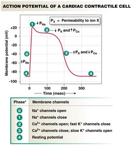
Contractile cells: Rapid depolarization via Na+ channels, brief repolarization, sustained plateau via L-type Ca2+ channels, final repolarization via K+ channels.
Plateau phase: Prolonged depolarization prevents summation and tetanus, ensuring rhythmic contractions.
Excitation-Contraction Coupling
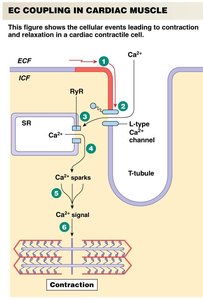
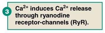
Cardiac muscle contraction is triggered by Ca2+ influx through L-type Ca2+ channels, which is not mechanically coupled to ryanodine receptors (RyR) as in skeletal muscle. Instead, Ca2+ entry induces further Ca2+ release from the sarcoplasmic reticulum (SR) via RyR, a process known as "Ca2+-induced Ca2+ release."
L-type Ca2+ channels: Allow extracellular Ca2+ entry.
RyR: Ryanodine receptors on SR release additional Ca2+.
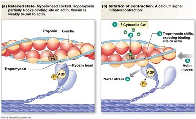
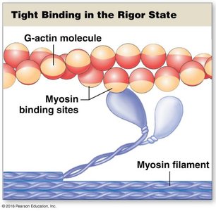
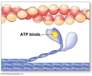
Cross-bridge cycling: Ca2+ binds to troponin, removing inhibition and allowing actin-myosin interaction.
Relaxation and Regulation
Relaxation occurs primarily via reuptake of Ca2+ into the SR, though removal to the extracellular space is also important. The SERCA pump (SR Ca2+-ATPase) is regulated by phospholamban (PLN); phosphorylation of PLN enhances Ca2+ uptake, increasing relaxation rates and contractility.
SERCA pump: Main mechanism for Ca2+ reuptake.
Phospholamban (PLN): Inhibits SERCA when dephosphorylated; phosphorylation removes inhibition.
Neuronal input: The autonomic nervous system modulates heart rate and contractility.
Sympathetic stimulation: Increases heart rate, conduction, contractility via phosphorylation of Ca2+ channels, RyR, SERCA, and myosin ATPase.
Parasympathetic stimulation: Decreases heart rate and conduction.
Force Generation and Length-Tension Relationship
Cardiac muscle is capable of graded single twitch contractions. The force of contraction is regulated by intracellular Ca2+ concentration and the length-tension relationship.
Graded contractions: Increased Ca2+ activates more troponin complexes, increasing cross-bridge formation.
Length-tension relationship: Slightly stretched sarcomeres generate greater force due to increased Ca2+ sensitivity and entry.
Feature | Skeletal Muscle | Cardiac Muscle |
|---|---|---|
Action Potential Duration | Short | Long (plateau phase) |
Summation/Tetanus | Possible | Prevented by long refractory period |
Ca2+ Source | SR only | Extracellular + SR |
Regulation | Neuronal | Autorhythmic + neuronal modulation |
Summary Table: Cardiac Muscle Physiology
Aspect | Cardiac Muscle |
|---|---|
Cell Type | Cardiac myocytes (contractile), autorhythmic cells |
Intercalated Disks | Desmosomes (mechanical), gap junctions (electrical) |
Action Potential | Long duration, plateau phase |
Ca2+ Handling | Ca2+-induced Ca2+ release |
Regulation | Autonomic nervous system, phospholamban |
Force Generation | Graded contractions, length-tension relationship |
Additional info: Academic context was added to clarify the mechanisms of excitation-contraction coupling, action potential phases, and regulatory pathways in cardiac muscle.
