 뒤로
Cardiac Physiology: Electrical Activity, Conduction, and Pumping
스터디 가이드 - 스마트 노트
 자료에 맞춘 맞춤형 노트, 핵심 정의, 예시, 맥락을 확장해 제공합니다.
Autonomic Control of the Heart
Sympathetic and Parasympathetic Pathways
The autonomic nervous system (ANS) modulates cardiac function through sympathetic and parasympathetic divisions, each with distinct pathways, neurotransmitters, and receptors. The heart's intrinsic pacemaker activity is modulated by these inputs to meet physiological demands.
Sympathetic Pathway: Increases heart rate, contractility, and AV nodal conduction via norepinephrine (NE) acting on β1-adrenergic receptors.
Parasympathetic Pathway: Primarily decreases heart rate and AV nodal conduction via acetylcholine (ACh) acting on M2 muscarinic receptors, with minimal direct effect on ventricular contractility.
Neural Pathway: CNS → preganglionic neuron (releases ACh at ganglion) → postganglionic neuron (releases NE or ACh at cardiac target) → cardiac tissue.
Key Terms:
Chronotropy: Rate of cardiac activity (heart rate). Positive chronotropy increases rate; negative decreases it.
Inotropy: Force of contraction. Positive inotropy increases force; negative decreases it.
Dromotropy: Speed of electrical conduction, especially through the AV node. Positive dromotropy increases conduction speed; negative decreases it.
Example: Increased vagal (parasympathetic) activity releases ACh at the SA and AV nodes, activating M2 receptors, which slows heart rate and AV conduction.
Cardiac Muscle and the Conduction System
Cardiac Cell Types and Connections
Cardiac muscle is specialized for rhythmic, involuntary contraction. Its structure supports coordinated pumping through both mechanical and electrical connections.
Striations: Organized contractile proteins, similar to skeletal muscle.
Branching Cells: Form a network for coordinated contraction.
Intercalated Discs: Specialized junctions containing:
Gap Junctions: Allow ion flow and electrical current between cells for rapid signal propagation.
Desmosomes: Provide mechanical strength to withstand contraction forces.
Functional Cell Types:
Autorhythmic (Pacemaker) Cells: Generate spontaneous action potentials (e.g., SA node, AV node).
Contractile Cardiomyocytes: Make up most of the myocardium; generate force in response to electrical activation.
Example: The SA node initiates the heartbeat, while contractile cells in the ventricles generate the force to eject blood.
Cardiac Conduction Pathway
The sequence of electrical activation ensures efficient pumping:
Sinoatrial (SA) Node: Dominant pacemaker in the right atrium; initiates each heartbeat.
Atrial Myocardium: Conducts activation through both atria.
Atrioventricular (AV) Node: Delays conduction, allowing ventricular filling.
AV Bundle (Bundle of His): Only normal electrical connection between atria and ventricles.
Right and Left Bundle Branches: Distribute activation to ventricles.
Purkinje Fibers: Rapidly conduct signals through ventricular myocardium.
Ventricular Myocardium: Contracts to eject blood.
Action Potentials in Cardiac Cells
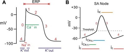
Pacemaker (SA Node) vs. Contractile Cell Action Potentials
Cardiac action potentials differ between pacemaker cells and contractile cardiomyocytes, reflecting their specialized roles.
Pacemaker Cells (SA Node):
Phase 4: Gradual depolarization (pacemaker potential) due to "funny" current (if), T-type Ca2+ channels, and reduced K+ efflux.
Phase 0: Depolarization via L-type Ca2+ channels.
Phase 3: Repolarization via K+ efflux.
Heart Rate Regulation: Slope of phase 4 determines rate; sympathetic input steepens slope (increases rate), parasympathetic flattens it (decreases rate).
Contractile Cells:
Phase 4: Stable resting potential.
Phase 0: Rapid Na+ influx (depolarization).
Phase 1: Brief repolarization (Na+ channels inactivate, K+ efflux).
Phase 2: Plateau (Ca2+ influx balances K+ efflux).
Phase 3: Repolarization (K+ efflux).
Excitation–Contraction Coupling in Cardiac Muscle
From Electrical Signal to Contraction
Electrical activation (action potential) triggers contraction through a process called excitation–contraction coupling, which relies on calcium dynamics.
Action Potential: Depolarizes the membrane and T-tubules.
Ca2+ Entry: L-type Ca2+ channels open, allowing extracellular Ca2+ influx.
Calcium-Induced Calcium Release (CICR): Influx triggers further Ca2+ release from the sarcoplasmic reticulum via ryanodine receptors.
Contraction: Increased cytosolic Ca2+ binds troponin, enabling actin–myosin cross-bridge cycling.
Relaxation: Ca2+ is pumped back into the SR (SERCA) and out of the cell (Na+/Ca2+ exchanger); Ca2+ dissociates from troponin, ending contraction.
Example: Sympathetic stimulation increases Ca2+ entry and SR cycling, enhancing contractile force (positive inotropy).
Electrocardiogram (ECG) and the Cardiac Cycle
ECG Waves and Intervals
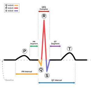
The ECG records the heart's electrical activity, with distinct waves corresponding to depolarization and repolarization events.
P wave: Atrial depolarization (precedes atrial contraction).
QRS complex: Ventricular depolarization (precedes ventricular contraction); atrial repolarization occurs but is masked.
T wave: Ventricular repolarization (precedes ventricular relaxation).
PR interval: Start of atrial depolarization to start of ventricular depolarization; includes AV nodal delay.
Cardiac Cycle Phases and Valve Function
The cardiac cycle consists of sequential phases, each defined by changes in pressure, volume, and valve state:
Ventricular Filling: AV valves open, blood flows into relaxed ventricle.
Atrial Systole: Atria contract, pushing additional blood into ventricle.
Isovolumetric Contraction: Both AV and semilunar valves closed; ventricular pressure rises, volume constant.
Ventricular Ejection: Semilunar valves open, blood ejected into arteries.
Isovolumetric Relaxation: Both valves closed; ventricular pressure falls, volume constant.
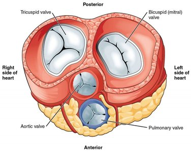
Valve Opening and Closing: Determined by pressure gradients, not direct muscle action.
Valve | Opens When... | Prevents Backflow From... |
|---|---|---|
Tricuspid | Right atrial pressure > right ventricular pressure | Right ventricle to right atrium |
Pulmonary | Right ventricular pressure > pulmonary artery pressure | Pulmonary artery to right ventricle |
Mitral (Bicuspid) | Left atrial pressure > left ventricular pressure | Left ventricle to left atrium |
Aortic | Left ventricular pressure > aortic pressure | Aorta to left ventricle |
Ventricular Volumes and Stroke Volume
End-Diastolic Volume (EDV): Volume in ventricle after filling (relaxation).
End-Systolic Volume (ESV): Volume remaining after contraction (not all blood is ejected).
Stroke Volume (SV): Amount ejected per beat.
Formula:
Heart Sounds
"Lub" (S1): Closure of AV valves (start of systole).
"Dub" (S2): Closure of semilunar valves (start of diastole).
Common Misconceptions and Clarifications
The ECG records electrical, not mechanical, events.
Valves open/close passively in response to pressure gradients, not direct muscle action.
Electrical activation precedes contraction; there is an electromechanical delay.
Not all blood is ejected from the ventricle with each beat.
Blood pressure measured in the arm is not the same as ventricular pressure.
Practice Questions
Which neurotransmitter is released by both sympathetic and parasympathetic preganglionic neurons?
Which receptor is activated by NE in the typical sympathetic pathway to the heart?
Define chronotropy, inotropy, and dromotropy.
What is the main functional difference between autorhythmic and contractile cardiomyocytes?
Which ECG feature represents ventricular depolarization?
What causes the aortic valve to open during a normal cardiac cycle?
Calculate stroke volume if EDV = 145 mL and ESV = 60 mL.
Additional info: For a comprehensive understanding, students should integrate knowledge of autonomic regulation, cardiac conduction, excitation–contraction coupling, and cardiac cycle mechanics to analyze physiological and clinical scenarios.
