 뒤로
Cardiac Physiology: Electrical Activity, Pressure Changes, and Heart Valves
스터디 가이드 - 스마트 노트
 자료에 맞춘 맞춤형 노트, 핵심 정의, 예시, 맥락을 확장해 제공합니다.
Cardiac Physiology: Electrical Activity to Pumping
Overview
This study guide covers the integration of electrical activation, muscle contraction, pressure changes, and valve function in the heart. It explains how these processes coordinate to move blood through the heart, focusing on the cardiac cycle, ECG interpretation, ventricular volumes, and heart sounds.
Electrocardiogram (ECG) and Electrical Events
ECG Waves and Their Significance
The ECG is a graphical representation of the heart's electrical activity, not a direct measure of mechanical contraction or pressure. Each wave corresponds to specific electrical events that precede mechanical actions in the heart.
P wave: Represents atrial depolarization, which precedes atrial contraction.
QRS complex: Represents ventricular depolarization, which precedes ventricular contraction. Atrial repolarization occurs here but is masked.
T wave: Represents ventricular repolarization, which precedes ventricular relaxation.
PR interval: Extends from the start of atrial depolarization to the start of ventricular depolarization, including AV nodal delay.
Mechanical contraction follows electrical activation with a short delay (electromechanical delay).
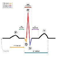
Additional info: The ECG does not provide information about chamber pressure, blood volume, or myocardial force.
Heart Valves and Pressure Gradients
Valve Function and Blood Flow
Heart valves are passive structures that open and close in response to pressure differences across them. They prevent backflow and direct blood flow through the heart.
Tricuspid valve: Opens when right atrial pressure exceeds right ventricular pressure; prevents backflow from right ventricle to right atrium.
Pulmonary valve: Opens when right ventricular pressure exceeds pulmonary artery pressure; prevents backflow from pulmonary artery to right ventricle.
Mitral valve: Opens when left atrial pressure exceeds left ventricular pressure; prevents backflow from left ventricle to left atrium.
Aortic valve: Opens when left ventricular pressure exceeds aortic pressure; prevents backflow from aorta to left ventricle.
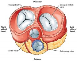
Additional info: Valves do not actively push blood; they respond to pressure gradients. Blood flows down a pressure gradient, and valves open or close accordingly.
Cardiac Cycle Phases
Sequence of Events
The cardiac cycle consists of a series of phases that coordinate blood movement through the heart. Each phase is defined by changes in pressure, valve state, and ventricular volume.
Ventricular filling: Ventricle is relaxed; AV valve opens as ventricular pressure is lower than atrial pressure; semilunar valve is closed.
Atrial systole: Atrial contraction increases atrial pressure, pushing more blood into the ventricle; AV valve open, semilunar valve closed.
Isovolumetric contraction: Ventricular contraction raises pressure; AV valve closes when ventricular pressure exceeds atrial pressure; semilunar valve still closed; both valves closed, volume constant.
Ventricular ejection: Semilunar valve opens when ventricular pressure exceeds arterial pressure; blood is ejected; AV valve remains closed.
Isovolumetric relaxation: Ventricle relaxes, pressure falls; semilunar valve closes when arterial pressure exceeds ventricular pressure; AV valve initially closed; both valves closed, volume constant until filling begins again.
Ventricular Volumes and Stroke Volume
Definitions and Calculation
Ventricular volumes change throughout the cardiac cycle, and these changes are used to calculate stroke volume.
End-Diastolic Volume (EDV): Volume in the ventricle after relaxation and filling.
End-Systolic Volume (ESV): Volume left in the ventricle at the end of contraction (ventricle does not empty completely).
Stroke Volume (SV): Amount of blood ejected per beat.
Formula:
Example: If EDV = 145 mL and ESV = 60 mL, then mL.
Heart Sounds
Origin and Clinical Significance
The "Lub" and "Dub" sounds heard via stethoscope are associated with valve closure:
Sound 1 "Lub": Closure of AV valves.
Sound 2 "Dub": Closure of semilunar valves.
These sounds result from vibrations of the valves, blood, and cardiac structures.
Clinicians listen for heart rate, rhythm, and murmurs (abnormal flow sounds indicating valve dysfunction).
Common Misconceptions in Cardiac Physiology
Clarifications
ECG waves represent electrical activation, not contraction.
ECG does not measure force, pressure, or blood volume.
Mechanical contraction follows electrical activation with a delay.
Valves open and close passively in response to pressure gradients.
AV and semilunar valves are not open simultaneously.
Blood moves down pressure gradients; valves prevent backflow.
During isovolumetric contraction, pressure increases while volume remains constant.
During isovolumetric relaxation, pressure decreases while volume remains constant.
Most ventricular filling is passive; atrial contraction adds the final portion.
Systole and diastole refer primarily to ventricular events; atrial and ventricular events overlap.
The ventricle does not empty completely with each beat.
Blood pressure readings reflect peripheral artery pressure, not ventricular pressure.
Practice Questions
Which ECG feature represents ventricular depolarization? Answer: B. QRS complex
What causes the aortic valve to open during a normal cardiac cycle? Answer: B. Ventricular pressure exceeds aortic pressure.
During isovolumetric contraction, which statement is correct? Answer: C. Both valves are closed and ventricular pressure rises.
A ventricle has an EDV of 145 mL and an ESV of 60 mL. Calculate stroke volume. Answer: SV = 145 mL - 60 mL = 85 mL
Explain why the ECG cannot tell us directly how much blood is ejected per beat. Answer: The ECG records electrical activity, not mechanical events or blood volume. Stroke volume is determined by changes in ventricular volume, not by electrical signals alone.
Heart Anatomy and Circulation
Chambers and Valves
The heart consists of four chambers (right atrium, right ventricle, left atrium, left ventricle) and four main valves (tricuspid, pulmonary, mitral, aortic) that regulate blood flow and prevent backflow.
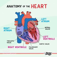
Additional info: Understanding the spatial arrangement of chambers and valves is essential for interpreting cardiac physiology and the effects of pressure changes.
