 뒤로
Cardiovascular System: Heart Anatomy, Physiology, and Blood Vessels
스터디 가이드 - 스마트 노트
 자료에 맞춘 맞춤형 노트, 핵심 정의, 예시, 맥락을 확장해 제공합니다.
Adrenal Gland Anatomy & Hormones
Adrenal Cortex and Medulla
The adrenal gland consists of two main regions: the cortex (outer) and medulla (inner), each producing distinct hormones crucial for homeostasis.
Aldosterone (Cortex): Increases sodium and water retention, raising blood pressure; promotes potassium excretion.
Cortisol (Cortex): Elevates blood glucose, mediates stress response, and acts as an anti-inflammatory.
Androgens (Cortex): Contribute to sex drive and development of pubic/axillary hair.
Epinephrine & Norepinephrine (Medulla): Initiate fight-or-flight response, increasing heart rate, blood pressure, and blood glucose.
Example: During acute stress, cortisol and epinephrine levels rise, preparing the body for rapid action.
Hypothalamus & Pituitary Gland Anatomy
Structural and Functional Connections
The hypothalamus connects to the pituitary gland via the infundibulum. It regulates hormone release through neural and vascular pathways.
Anterior Pituitary: Controlled by hypothalamic hormones via hypophyseal portal vessels.
Posterior Pituitary: Connected by axons; stores/releases ADH and oxytocin produced by the hypothalamus.
Example: ADH release increases water retention in kidneys during dehydration.
Hormone Functional Classifications
Types of Hormonal Actions
Hormones are classified based on their effects and interactions with other hormones.
Tropic: Stimulate other endocrine glands (e.g., TSH, ACTH).
Non-tropic: Act directly on target tissues (e.g., insulin, ADH).
Synergistic: Work together to amplify an effect.
Antagonistic: Oppose each other's actions (e.g., insulin vs. glucagon).
Permissive: One hormone enhances another’s effect.
Calcium Homeostasis Hormones
Regulation of Blood Calcium Levels
Calcium homeostasis is maintained by hormones from the parathyroid and thyroid glands, as well as vitamin D.
PTH (Parathyroid): Increases blood calcium by stimulating bone resorption, kidney retention, and vitamin D activation.
Calcitonin (Thyroid): Decreases blood calcium by inhibiting osteoclasts.
Calcitriol (Active Vitamin D): Increases intestinal calcium absorption.
Example: Low blood calcium triggers PTH release; high calcium triggers calcitonin (minor role in adults).
Thymus Gland & Immune Function
Location and Hormones
The thymus, located behind the sternum, produces hormones that promote T-cell maturation, essential for adaptive immunity.
Thymosin, thymopoietin, thymulin: Support T-cell development.
Blood Glucose Homeostasis (Pancreas)
Insulin and Glucagon Regulation
The pancreas regulates blood glucose through insulin and glucagon, maintaining homeostasis via negative feedback.
Insulin (β cells): Lowers blood glucose by increasing cellular uptake and glycogen storage.
Glucagon (α cells): Raises blood glucose by stimulating glycogen breakdown and glucose production in the liver.
Example: After a meal, insulin is released to reduce blood glucose; during fasting, glucagon increases blood glucose.
Heart Anatomy & Conduction System
Chamber Structure and Blood Flow
The heart consists of four chambers and four valves, ensuring unidirectional blood flow and efficient circulation.
Chambers: Right/left atria (receive blood); right/left ventricles (pump blood).
Valves: Tricuspid (RA→RV), pulmonary (RV→lungs), mitral/bicuspid (LA→LV), aortic (LV→body).
Interventricular Septum: Separates right and left sides.
Blood Flow Pathway: Body → SVC/IVC → RA → Tricuspid → RV → Pulmonary Valve → Pulmonary Arteries → Lungs → Pulmonary Veins → LA → Mitral → LV → Aortic Valve → Aorta → Body.
Electrical Conduction Pathway
The heart’s electrical system coordinates contraction and relaxation for effective pumping.
SA node: Natural pacemaker; initiates heartbeat.
AV node: Delays impulse for ventricular filling.
Bundle of His → R/L bundle branches → Purkinje fibers: Conduct impulses for ventricular contraction.
Heart Wall Anatomy
Layers and Functions
The heart wall is composed of three layers, each with distinct functions.
Epicardium: Outer protective layer; visceral layer of serous pericardium.
Myocardium: Middle layer; cardiac muscle responsible for contraction.
Endocardium: Inner smooth lining of chambers and valves.
Pericardium: Protective sac; pericardial fluid reduces friction.
Coronary Circulation
Heart’s Blood Supply
Coronary arteries supply oxygenated blood to the myocardium, while cardiac veins return deoxygenated blood to the right atrium.
Right Coronary Artery (RCA): Supplies right heart; branches into right marginal and posterior interventricular arteries.
Left Coronary Artery (LCA): Branches into LAD (anterior interventricular) and circumflex arteries.
Cardiac Veins → Coronary Sinus → Right Atrium: Return deoxygenated blood.
Example: Most coronary blood flow occurs during ventricular diastole.
Cardiac Cycle: Systole and Diastole
Phases of the Cardiac Cycle
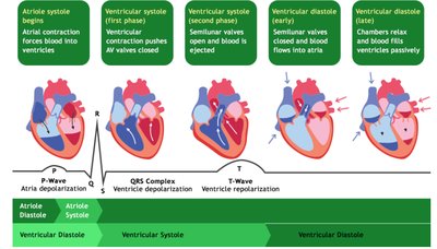
The cardiac cycle consists of alternating periods of contraction (systole) and relaxation (diastole), enabling blood flow through the heart.
Systole: Contraction phase; blood is pumped out of the chambers.
Diastole: Relaxation phase; chambers fill with blood.
Example: During ventricular systole, blood is ejected into the arteries; during diastole, chambers refill.
Cardiac Output (CO) and Stroke Volume (SV)
Formulas and Laws
Cardiac output and stroke volume are key measures of heart function, influenced by heart rate, contractility, and preload.
Cardiac Output (CO): Blood pumped by each ventricle per minute.
Formula:
Stroke Volume (SV): Blood ejected per beat.
Formula:
Frank-Starling Law: Increased venous return increases EDV and SV.
Contractility: Stronger contraction decreases ESV, increasing SV.
Afterload: Increased resistance increases ESV, decreasing SV.
Example: Normal resting CO is ~5 L/min; normal SV is ~70 mL/beat.
ECG Peaks, Segments & Intervals
Electrocardiogram Interpretation
The ECG records electrical activity of the heart, with distinct waves and intervals corresponding to cardiac events.
P wave: Atrial depolarization.
QRS complex: Ventricular depolarization (atrial repolarization hidden).
T wave: Ventricular repolarization.
PR segment: AV node conduction delay.
ST segment: Ventricles fully depolarized.
PR interval: Start P → start QRS; atrial-to-ventricular conduction (0.12–0.20 sec).
QT interval: Start QRS → end T; ventricular depolarization + repolarization.
QRS duration: Ventricular depolarization (normally <0.12 sec).
Example: Normal sinus rhythm: 60–100 bpm, regular R-R intervals, one P wave before every QRS.
Heart Sounds
Phases and Clinical Significance
Heart sounds are produced by valve closures and blood flow, with clinical relevance for diagnosing cardiac conditions.
S1: AV valves close; start of ventricular systole.
S2: Semilunar valves close; start of ventricular diastole.
S3: Rapid ventricular filling; normal in youth, may indicate heart failure in adults.
S4: Atrial contraction against stiff ventricle; usually abnormal.
Murmur: Turbulent blood flow, often from valve stenosis or regurgitation.
Blood Vessel Anatomy
Structure and Types
Blood vessels are classified by structure and function, facilitating circulation and exchange.
Tunica Intima: Inner layer; endothelium reduces friction.
Tunica Media: Middle layer; smooth muscle for vasoconstriction/vasodilation.
Tunica Externa: Outer layer; connective tissue for protection/support.
Arteries: Carry blood away from heart; thick, elastic walls.
Arterioles: Control blood flow and BP.
Capillaries: One-cell-thick walls; exchange of gases, nutrients, and wastes.
Venules: Collect blood from capillaries.
Veins: Return blood to heart; thin walls, valves prevent backflow.
Blood Pressure (BP) and Regulation
Forces, Components, and Regulation
Blood pressure is determined by cardiac output and peripheral resistance, regulated by neural and hormonal mechanisms.
Systolic BP: Highest pressure during ventricular contraction.
Diastolic BP: Lowest pressure during ventricular relaxation.
Pulse Pressure (PP): Systolic − Diastolic.
Mean Arterial Pressure (MAP): Average arterial pressure; drives tissue perfusion.
Formula:
BP Formula:
Example: 120/80 mmHg → PP = 40; MAP ≈ 93 mmHg.
Capillary Types and Permeability
Classification and Function
Capillaries are classified by permeability, affecting exchange of substances.
Continuous: Least permeable; tight junctions. Found in muscles, skin, lungs, brain (BBB).
Fenestrated: Small pores; rapid filtration/absorption. Found in kidneys, intestines, endocrine glands.
Sinusoidal: Large gaps; most permeable. Found in liver, spleen, bone marrow.
Blood Pressure Regulation Mechanisms
Neural and Hormonal Control
Blood pressure is regulated by short-term neural and long-term hormonal/kidney mechanisms.
Neural: Baroreceptors detect BP changes; medulla adjusts SNS/PSNS, HR, and vessel diameter.
RAAS: Low BP triggers renin → angiotensin II (vasoconstriction) → aldosterone (increases Na⁺/water retention).
ADH: Increases water retention and vasoconstriction.
ANP: Increases Na⁺/water excretion.
Kidneys: Regulate blood volume via urine output.
Chemoreceptors: Detect low O₂, high CO₂, low pH; stimulate SNS.
Capillary Net Filtration (Starling Forces)
Fluid Movement Across Capillaries
Starling forces determine net fluid movement across capillary walls, balancing hydrostatic and osmotic pressures.
Blood Hydrostatic Pressure (BHP): Pushes fluid out of capillaries.
Blood Colloid Osmotic Pressure (BCOP): Plasma proteins pull fluid in.
Interstitial Hydrostatic Pressure (IFHP): Pushes fluid in.
Interstitial Colloid Osmotic Pressure (IFCOP): Pulls fluid out.
Net Filtration Pressure (NFP):
Example: Positive NFP = filtration (fluid out); negative NFP = reabsorption (fluid in).
Major Arteries and Veins of the Upper Limb
Blood Flow Pathways
Arteries and veins of the upper limb follow a specific pathway, ensuring efficient circulation.
Arteries: Subclavian → Axillary → Brachial → Radial & Ulnar → Palmar Arches → Digital Arteries.
Veins: Radial/Ulnar → Brachial → Axillary → Subclavian; superficial veins include cephalic, basilic, median cubital.
Case Study: Thyroid Disorders
Hormone Pathway and Symptoms
Thyroid disorders affect metabolism and are diagnosed by hormone levels and clinical symptoms.
Pathway: Hypothalamus (TRH) → Anterior Pituitary (TSH) → Thyroid (T3/T4).
Negative Feedback: Increased T3/T4 decreases TRH & TSH; decreased T3/T4 increases TRH & TSH.
Hypothyroidism: Fatigue, weight gain, cold intolerance, slow HR.
Hyperthyroidism: Weight loss, heat intolerance, rapid HR, anxiety.
Disorder | T3/T4 | TSH |
|---|---|---|
Primary Hypothyroidism | ↓ | ↑ |
Primary Hyperthyroidism | ↑ | ↓ |
Secondary Hypothyroidism (pituitary) | ↓ | ↓ |
Secondary Hyperthyroidism (pituitary) | ↑ | ↑ |
Case Study: Ventricular Septal Defect (VSD)
Pathophysiology and ECG Changes
VSD is a congenital heart defect causing abnormal blood flow and potential cardiac complications.
VSD: Hole in interventricular septum; blood leaks from LV to RV (left-to-right shunt).
Effects: Increased pulmonary flow, increased RV workload, decreased effective systemic SV.
ECG Changes: Small VSD often normal; large VSD may show RV/LV hypertrophy, increased QRS voltage, possible axis deviation.
Example: Severe VSD can lead to ventricular enlargement and heart failure.
Additional info: Academic context was added to expand brief points and ensure completeness for exam preparation.
