 뒤로
Ch.12 Part 3
스터디 가이드 - 스마트 노트
 자료에 맞춘 맞춤형 노트, 핵심 정의, 예시, 맥락을 확장해 제공합니다.
The Central Nervous System
Functional Brain Systems
The central nervous system (CNS) contains networks of neurons that span wide areas of the brain, forming functional systems. Two major systems are the limbic system and the reticular formation.
Limbic System
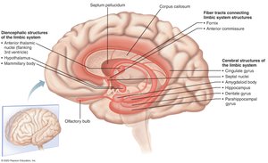
The limbic system is a group of structures located on the medial aspects of the cerebral hemispheres and diencephalon. It is involved in emotional responses, memory, and motivation.
Fornix: A fiber tract linking limbic system regions.
Amygdaloid body: Recognizes angry or fearful facial expressions, assesses danger, and elicits fear responses.
Cingulate gyrus: Expresses emotions via gestures and resolves mental conflict.
Hippocampus: Plays a role in memory formation and retrieval.
Most output is relayed via the hypothalamus, which is involved in psychosomatic illnesses.
The limbic system interacts with the prefrontal lobes, allowing emotional reactions to conscious understanding and awareness of emotional richness.
Reticular Formation
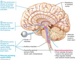
The reticular formation extends through the central core of the brain stem and consists of three broad columns of nuclei. It has widespread axonal connections, allowing it to govern brain arousal and consciousness.
Reticular activating system (RAS): Sends impulses to the cerebral cortex to keep it conscious and alert; filters out repetitive or weak stimuli.
Inhibited by sleep centers, alcohol, and drugs.
Severe injury can result in permanent unconsciousness (coma).
Motor function helps control coarse limb movements via reticulospinal tracts.
Autonomic centers regulate visceral motor functions (vasomotor, cardiac, respiratory centers).
Higher Mental Functions
Language
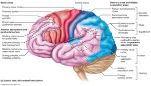
Language processing involves the association cortex of the left hemisphere. Two main areas are:
Broca’s area: Responsible for speech production. Lesions cause inability to speak, though comprehension remains intact.
Wernicke’s area: Responsible for understanding spoken and written words. Lesions cause nonsensical speech, though the ability to speak is preserved.
Corresponding areas on the right hemisphere handle nonverbal language components.
Memory
Memory is the storage and retrieval of information. It is classified into several types:
Declarative memory: Facts (names, faces, words, dates).
Procedural memory: Skills (e.g., playing piano).
Motor memory: Motor skills (e.g., riding a bike).
Emotional memory: Experiences linked to emotions (e.g., heart pounding when hearing a rattlesnake).
Declarative memory storage occurs in two stages:
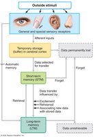
Short-term memory (STM): Temporary holding of information, limited to 7–8 items.
Long-term memory (LTM): Limitless capacity for information storage.
Factors affecting transfer from STM to LTM include emotional state, rehearsal, association, and automatic memory. Memory consolidation involves fitting new facts into categories already stored in the cerebral cortex, with the hippocampus, temporal cortical areas, thalamus, and prefrontal cortex involved.
Clinical Memory Disorders
Anterograde amnesia: Inability to form new memories; old memories remain intact.
Retrograde amnesia: Loss of memories formed in the distant past.
Brain Wave Patterns and EEG
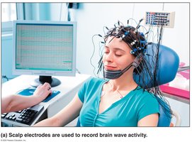
Brain waves reflect electrical activity of higher mental functions and are measured by an electroencephalogram (EEG). EEG is used to diagnose epilepsy, sleep disorders, and to determine brain death.
Electrodes placed on the scalp measure electrical potential differences between cortical areas.
Brain waves are unique to each person and change with age, sensory stimuli, brain disease, and chemical state.
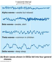
Wave frequency is measured in Hertz (Hz), with four main classes:
Alpha waves (8–13 Hz): Regular, rhythmic, low-amplitude; indicate an "idling" brain.
Beta waves (14–30 Hz): Rhythmic, less regular; occur when mentally alert.
Theta waves (4–7 Hz): More irregular; common in children, uncommon in awake adults.
Delta waves (≤4 Hz): High-amplitude; deep sleep or brain damage in awake adults.
Sleep and Sleep-Wake Cycles
Types and Stages of Sleep
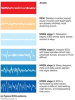
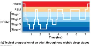
Sleep is a state of partial unconsciousness from which a person can be aroused. Two major types are defined by EEG patterns:
Non–rapid eye movement (NREM) sleep: Four stages, with frequency of waves declining and amplitude increasing.
Rapid eye movement (REM) sleep: Begins abruptly after ~90 minutes; temporary paralysis except for eye movements; most dreaming occurs in REM.
Sleep Regulation and Importance
Sleep and wakefulness reflect natural circadian rhythms.
RAS activity is inhibited during sleep but mediates sleep stages.
Hypothalamus releases orexins to help cortex wake up.
Slow-wave sleep (NREM stages 3 and 4) is restorative.
REM sleep may help analyze and process emotional events, and eliminate unneeded synapses.
Daily sleep requirements decline with age; stage 4 sleep may disappear after age 60.
Clinical Sleep Disorders
Narcolepsy: Abrupt lapse into sleep from awake state; orexins destroyed by immune system.
Cataplexy: Loss of muscle tone while awake.
Insomnia: Chronic inability to obtain sufficient sleep; may be treated by blocking orexin action.
Consciousness
Levels and Clinical Disorders
Consciousness involves perception, voluntary movement, and higher mental processing. It is clinically graded as alertness, drowsiness, stupor, and coma.
Loss of consciousness signals impaired brain function.
Fainting (syncope): Brief loss of consciousness, often due to inadequate cerebral blood flow.
Coma: Extended unconsciousness; oxygen consumption is lowered.
Brain death: Irreversible coma; ethical and legal issues arise.
Clinical Disorders: Epilepsy
Epileptic seizures are caused by abnormal electrical discharges in groups of neurons, preventing normal messages from getting through.
Epilepsy is not associated with intellectual impairments; occurs in 1% of the population.
Causes include genetic factors, brain injuries, stroke, infections, or tumors.
Aura: Sensory hallucination preceding a seizure.
Absence seizures: Mild, brief blank expression; common in children.
Tonic-clonic seizures: Severe, lasting minutes; loss of consciousness, incontinence, severe biting of tongue.
Post-ictal state: Confusion and memory loss immediately after a seizure.
Cerebrospinal Fluid (CSF)
Formation and Function
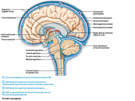
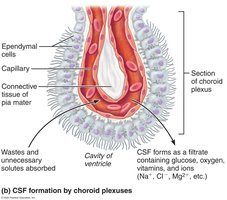
CSF forms a liquid cushion around the brain, providing buoyancy, protection, and nourishment. It is produced by the choroid plexus, a cluster of capillaries enclosed by pia mater and ependymal cells.
Reduces brain weight by 97%.
Protects CNS from trauma.
Nourishes brain and carries chemical signals.
Composed of a watery solution from blood plasma, with less protein and different ion concentrations.
Normal adult CSF volume is ~150 mL, replaced every 8 hours.
Blood-Brain Barrier
Structure and Function
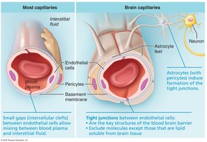
The blood-brain barrier maintains a stable environment for the brain by restricting passage of substances from blood to neurons.
Substances must pass through continuous endothelium of capillary walls.
Tight junctions ensure substances pass through, not around endothelial cells.
Astrocyte feet and pericytes promote tight junction formation.
Substances move via simple diffusion, facilitated diffusion, or transcytosis.
Thick basement membrane contains enzymes that destroy chemicals activating neurons.
Barrier is absent in some areas (vomiting center, hypothalamus) to monitor blood composition and temperature.
Brain Injuries and Disorders
Traumatic Brain Injuries
Concussion: Temporary alteration in function.
Contusion: Permanent damage.
Subdural/subarachnoid hemorrhage: Pressure from blood may force brain stem through foramen magnum, resulting in death.
Cerebral edema: Swelling associated with traumatic head injury.
Cerebrovascular Accidents (CVAs)
Also called "strokes."
Ischemia: Tissue deprived of blood supply, leading to death of brain tissue; often caused by blockage of cerebral artery.
Glutamate acts as excitotoxin, worsening condition.
Hemiplegia, sensory, and speech deficits may result.
Transient ischemic attacks (TIAs): Temporary, reversible episodes.
Tissue plasminogen activator (TPA): Approved treatment for stroke.
Degenerative Brain Disorders
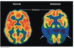
Alzheimer’s disease (AD): Progressive degenerative disease causing dementia.
Key proteins misfold and malfunction.
Symptoms: memory loss, short attention span, disorientation, language loss, irritability, moodiness, confusion, hallucinations.
Plaques of beta-amyloid peptides form in the brain.
Neurofibrillary tangles inside neurons interfere with transport, eventually killing neurons.
As brain cells die, the brain shrinks.
Additional info: The notes above expand on brief points with academic context, definitions, and examples, and include only images that are directly relevant to the explanation of each paragraph.
