 뒤로
Central Nervous System: Structure and Function
스터디 가이드 - 스마트 노트
 자료에 맞춘 맞춤형 노트, 핵심 정의, 예시, 맥락을 확장해 제공합니다.
The Central Nervous System
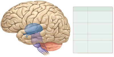
Overview of Brain Divisions
The central nervous system (CNS) consists of the brain and spinal cord, which are responsible for processing and integrating information, maintaining homeostasis, and controlling movement and reflexes. The adult human brain contains 97% of the body's nervous tissue.
Cerebrum: Performs higher mental functions, interprets sensory stimuli, plans and initiates movement.
Diencephalon: Processes, integrates, and relays information; maintains homeostasis; regulates biological rhythms.
Cerebellum: Monitors and coordinates movement; maintains homeostasis.
Brainstem: Controls certain reflexes; monitors movement; integrates and relays information.
Spinal Cord: Relays information between the brain and peripheral nervous system.
Physical Protection of the Brain
The brain is protected by several structures:
Bones of the cranium: Provide rigid protection.
Cranial meninges: Three layers (dura mater, arachnoid mater, pia mater) surround the brain.
Cerebrospinal fluid (CSF): Cushions and nourishes the CNS.
Blood-brain barrier: Astrocytes regulate passage of substances from blood to brain tissue.
Gray Matter vs. White Matter
Distribution in Brain and Spinal Cord
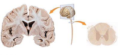
Gray matter and white matter are distributed differently in the brain and spinal cord.
Gray matter: Composed mostly of cell bodies; lacks myelination. In the brain, it forms the outer cortex; in the spinal cord, it is central.
White matter: Composed mostly of myelinated axons. In the brain, it is deep to the cortex; in the spinal cord, it is peripheral.
Brain Ventricles and Cerebrospinal Fluid (CSF)
Structure and Flow of CSF
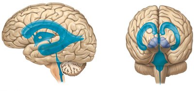
The brain contains interconnected ventricles filled with CSF, which is produced by choroid plexuses and circulates to provide nutrients, buoyancy, and cushioning.
Lateral ventricles: Located in each cerebral hemisphere.
Third ventricle: Located in the diencephalon.
Fourth ventricle: Located between the brainstem and cerebellum.
CSF flow: CSF is secreted by ependymal cells, circulates through ventricles and subarachnoid space, and is reabsorbed into blood via dural sinuses.
Cerebral Lobes and Functional Areas
Lobes of the Cerebrum
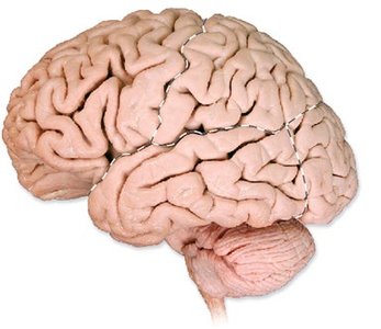
The cerebrum is divided into four main lobes, each with distinct functions:
Frontal lobe: Voluntary motor function, motivation, mood, personality, decision making.
Parietal lobe: Reception and evaluation of sensory information and taste.
Occipital lobe: Visual input processing and integration.
Temporal lobe: Hearing, smell, memory, abstract thought, judgment.
Gyri and Sulci
Gyri: Ridges on the cerebral surface.
Sulci: Grooves separating gyri.
Central sulcus: Separates precentral gyrus (primary motor cortex) and postcentral gyrus (primary somatosensory cortex).
Functional Mapping: Motor and Sensory Homunculi
Motor and Sensory Cortex
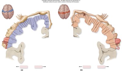
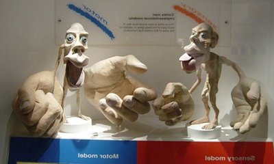
The precentral gyrus contains the primary motor cortex, while the postcentral gyrus contains the primary somatosensory cortex. These areas are mapped to different body regions, forming the motor and sensory homunculi.
Motor homunculus: Represents the distribution of motor control.
Sensory homunculus: Represents the distribution of sensory input.
Cerebral White Matter: Tracts and Fibers
Types of White Matter Tracts
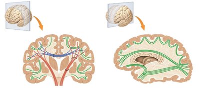
White matter in the cerebrum is organized into three main types of tracts:
Association tracts: Connect gyri within the same hemisphere.
Commissural tracts: Connect corresponding regions in opposite hemispheres (e.g., corpus callosum).
Projection tracts: Link the cerebral cortex to lower brain regions and spinal cord.
Basal Nuclei and Limbic System
Basal Nuclei
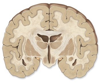
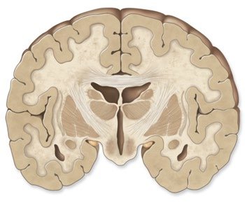
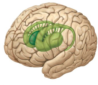
Basal nuclei are deep gray matter structures involved in motor control.
Caudate nucleus: Produces pattern and rhythm of walking movements.
Putamen: Controls movements at a subconscious level.
Globus pallidus: Adjusts muscle tone via thalamus.
Limbic System
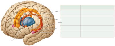
The limbic system is a functional unit involved in emotion, memory, and survival behaviors.
Structures: Includes cingulate gyri, parahippocampal gyrus, hippocampus, amygdala, and fornix.
Functions: Establishes emotional states, links conscious and autonomic functions, and supports memory and learning.
Cerebral Lateralization
Hemispheric Specialization
The left and right hemispheres of the cerebrum are specialized for different functions.
Left hemisphere: Language, mathematics, motor speech (Broca area), verbal memory.
Right hemisphere: Spatial relationships, music ability, recognition of faces, memory for shapes.
Lateralization: Develops in early childhood and is correlated with handedness.
Diencephalon and Brainstem
Diencephalon Structures and Functions
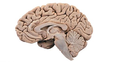
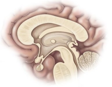
The diencephalon includes the thalamus, hypothalamus, and epithalamus.
Thalamus: Relays and filters sensory information to the cerebrum.
Hypothalamus: Maintains homeostasis, controls autonomic and endocrine systems, regulates emotional behavior, body temperature, food and water intake, and sleep-wake rhythms.
Epithalamus: Contains the pineal gland, which regulates circadian rhythms and initiates puberty.
Brainstem Functions
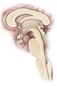
The brainstem consists of the midbrain, pons, and medulla oblongata.
Midbrain: Processes sensory data (except olfaction), controls reflexive somatic motor responses.
Pons: Relays sensory information, regulates sleep and breathing.
Medulla oblongata: Regulates visceral functions such as heart rate, blood vessel diameter, respiration, swallowing, and reflexes.
Reticular Formation
The reticular formation is a group of nuclei scattered throughout the brainstem, playing roles in arousal, awareness, posture, pain, sleep-wake cycle, alertness, consciousness, focusing, and attention.
Cerebellum
Structure and Function
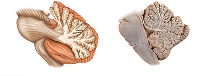
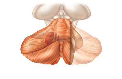
The cerebellum is responsible for adjusting postural muscles, governing balance, regulating conscious movements, and learning motor skills.
Cerebellar cortex: Outer layer of gray matter.
Arbor vitae: Inner white matter resembling a tree.
Deep cerebellar nuclei: Embedded within the white matter.
Motor Control and Integration
Modifying and Refining Motor Activities
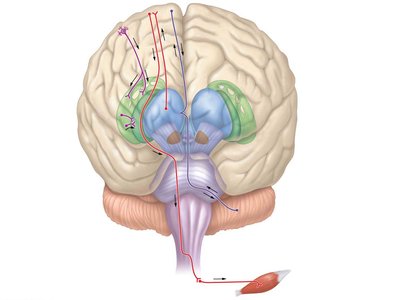
Motor activities are planned and refined by feedback loops among basal nuclei, thalamus, and cerebral cortex.
Basal nuclei: Facilitate or inhibit muscle activity, coordinate movements and posture.
Cerebellum: Maintains muscle tone, controls balance, coordinates eye movements.
Cranial Meninges and Dural Sinuses
Structure and Function
The cranial meninges consist of two layers of dura mater (periosteal and meningeal), arachnoid mater, and pia mater. Dural folds (falx cerebri, tentorium cerebelli, falx cerebelli) and venous sinuses provide structural support and drainage. 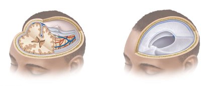
Spinal Cord Structure
Spinal Cord and Meninges
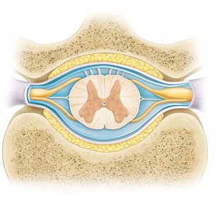
The spinal cord is protected by three meninges: dura mater, arachnoid mater, and pia mater. It contains enlargements, conus medullaris, cauda equina, and filum terminale. 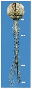
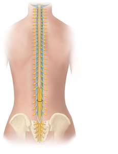
Spinal Cord Gray and White Matter
Gray matter: Contains cell bodies; divided into anterior, posterior, and lateral horns.
White matter: Contains myelinated axons; divided into funiculi.
Central canal: Contains CSF.
Ascending and Descending Tracts
Tracts of the Spinal Cord
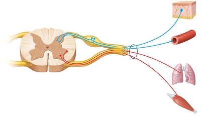
Ascending tracts: Carry sensory information to the brain (e.g., posterior columns, spinocerebellar, spinothalamic tracts).
Descending tracts: Carry motor information from the brain to the body (e.g., corticospinal, reticulospinal, tectospinal, vestibulospinal tracts).
Ascending Tract | Function |
|---|---|
Posterior columns—fasciculus gracilis | Fine touch, vibration, proprioception from lower limbs |
Posterior columns—fasciculus cuneatus | Fine touch, vibration, proprioception from trunk, neck, upper limbs |
Spinocerebellar tracts | Proprioceptive information to cerebellum |
Anterolateral system—spinothalamic tracts | Pain, temperature, certain types of touch |
Descending Tract | Function |
|---|---|
Corticospinal tracts | Motor information from cerebral cortex |
Reticulospinal tracts | Motor information from brainstem; posture |
Tectospinal tract | Reflexive movement of head and eyes |
Vestibulospinal tract | Posture and balance |
Higher Order Brain Functions
EEG, Speech, Memory, and Sleep
Higher order brain functions require the cerebral cortex and involve both conscious and unconscious processing.
EEG: Measures electrical activity (brain waves: alpha, beta, theta, delta).
Speech: Wernicke area (understanding), Broca area (production).
Memory: Immediate, short-term, and long-term memory; consolidation enhanced by association and emotion.
Sleep: Alternates between REM and non-REM stages; REM sleep involves active dreaming and decreased muscle tone.
Representative Diseases and Disorders of the CNS
Movement Disorders
Parkinson disease: Lesion in basal nuclei; rigidity, tremor, slow movement.
Dyskinesias: General movement disorders involving basal nuclei.
Huntington disease: Hereditary degeneration of basal nuclei; involuntary movements.
Cerebral palsy: Motor defects due to brain damage; increased tone, tremors, difficulty speaking/swallowing.
Cerebellar lesions: Ataxic, jerky movements; intention tremor.
Other CNS Disorders
Alzheimer disease: Dementia, loss of neurons in cerebral cortex; memory loss, disorientation.
Tay-Sachs disease: Hereditary lipid-storage disorder; paralysis, blindness, death.
Epilepsy: Seizures from massive neuronal discharge.
Headaches: Various causes including inflammation and tension.
Dyslexia: Reading deficiency; confusion between similar letters.
Stroke: Bleeding or blockage in brain; loss of speech, numbness, paralysis.
