 뒤로
Chapter 10: The Muscular System – Study Guide for Anatomy & Physiology
스터디 가이드 - 스마트 노트
 자료에 맞춘 맞춤형 노트, 핵심 정의, 예시, 맥락을 확장해 제공합니다.
Chapter 10: The Muscular System
10.1 Muscle Actions and Interactions
The muscular system is responsible for movement and stability in the human body. Muscles act by pulling, never pushing, and their actions are determined by their position relative to the joints they cross. There are three main functional groups of muscles:
Prime mover (agonist): The muscle primarily responsible for a specific movement.
Antagonist: Opposes or reverses the action of the prime mover.
Synergist: Assists the prime mover by adding force or reducing unnecessary movement.
Fixator: A type of synergist that stabilizes the origin of the prime mover.
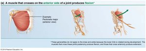
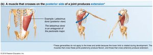
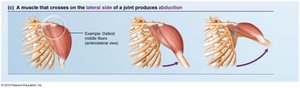
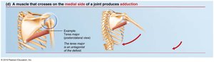
Muscle action can be inferred by the position of the muscle relative to the joint it crosses:
Anterior side: produces flexion
Posterior side: produces extension
Lateral side: produces abduction
Medial side: produces adduction
10.2 Naming Skeletal Muscles
Skeletal muscles are named based on several criteria, which often describe their characteristics or location:
Location: Associated bone or region (e.g., temporalis over temporal bone).
Shape: Distinctive shape (e.g., deltoid = triangle).
Size: Maximus (largest), minimus (smallest), longus (long).
Direction of fibers: Rectus (straight), transversus (right angles), oblique (angles).
Number of origins: Biceps (2), triceps (3).
Location of attachments: Origin and insertion (e.g., sternocleidomastoid).
Action: Flexor, extensor, adductor, etc.
Often, several criteria are combined in a muscle's name (e.g., extensor carpi radialis longus).
10.3 Fascicle Arrangements
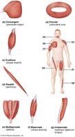
Fascicles are bundles of muscle fibers, and their arrangement determines the muscle's shape and functional capabilities. The most common patterns are:
Circular: Fascicles arranged in concentric rings (e.g., orbicularis oris).
Convergent: Broad origin, fascicles converge toward a single tendon (e.g., pectoralis major).
Parallel: Fascicles parallel to the long axis (e.g., sartorius).
Fusiform: Spindle-shaped with parallel fibers (e.g., biceps brachii).
Pennate: Short fascicles attach obliquely to a central tendon (e.g., rectus femoris).
Pennate muscles can be unipennate, bipennate, or multipennate, depending on the arrangement of fascicles.
10.4 Lever Systems
Most skeletal muscles move bones using lever systems. A lever consists of:
Lever: Rigid bar (bone)
Fulcrum: Fixed point (joint)
Effort: Force applied by muscle contraction
Load: Resistance moved by the effort
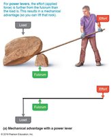
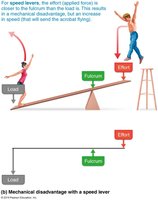
Levers operate at either mechanical advantage (power lever) or mechanical disadvantage (speed lever):
Mechanical advantage: Load is close to fulcrum, effort is far; small effort moves large load.
Mechanical disadvantage: Load is far from fulcrum, effort is close; load moves rapidly over large distance.
Classes of Levers
Levers are classified based on the relative positions of effort, fulcrum, and load:
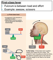
First-class lever: Fulcrum is between load and effort (e.g., seesaw, scissors, raising head). Can be mechanical advantage or disadvantage.
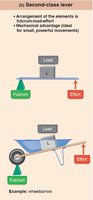
Second-class lever: Load is between fulcrum and effort (e.g., wheelbarrow, standing on toes). Always mechanical advantage.
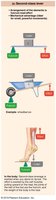
Third-class lever: Effort is between fulcrum and load (e.g., tweezers, most skeletal muscles). Always mechanical disadvantage.
Equation for lever mechanics:
10.5 Major Skeletal Muscles of the Body
There are over 600 skeletal muscles, grouped by function and location. Muscle tables typically include:
Description: Location relative to other muscles
Origin and insertion: Usually a joint between origin and insertion
Actions: Movement caused by contraction
Innervation: Major nerve supplying the muscle
Tips for learning muscles:
Use muscle names to infer location and function
Identify muscles on anatomical figures
Relate location and attachments to actions
Act out movements and palpate muscles
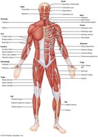
Muscles of the Head and Neck
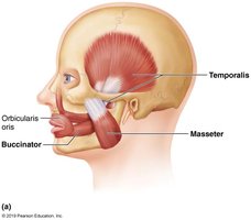
Muscles of facial expression insert into skin, not bone, and are important for nonverbal communication. All are innervated by cranial nerve VII (facial nerve). Groups include:
Muscles of the scalp
Muscles of the face
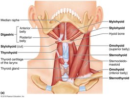
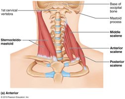
Muscles of the Neck and Vertebral Column
These muscles move the head and trunk, including the sternocleidomastoid and scalene muscles. They are essential for head movement and posture.
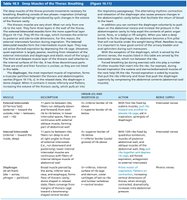
Deep Muscles of the Thorax: Breathing
Breathing involves two phases: inspiration and expiration. Inspiratory muscles include the diaphragm and external intercostals, which enlarge the rib cage. Expiration is brought on by relaxation of inspiratory muscles and contraction of internal intercostals.
Muscles of the Abdominal Wall
The abdominal wall is formed by several muscles, including the rectus abdominis, external and internal obliques, and transversus abdominis. These muscles support the abdominal organs and aid in movements such as flexion and rotation of the trunk.
Muscles of the Shoulder and Arm
Muscles acting on the scapula and arm include the deltoid, pectoralis major, latissimus dorsi, and rotator cuff muscles. Their actions include elevation, depression, rotation, and abduction/adduction of the arm.
Muscles Crossing the Elbow Joint
Muscles crossing the elbow joint are divided into anterior (flexors) and posterior (extensors) compartments. Examples include biceps brachii (flexor) and triceps brachii (extensor).
Muscles Crossing the Hip and Knee Joints
Muscles are grouped as anterior, medial, or posterior. Anterior muscles flex the femur and extend the leg; posterior muscles extend the thigh and flex the leg; medial muscles adduct the thigh. The quadriceps femoris is a powerful knee extensor, while the hamstrings are prime movers of thigh extension and knee flexion.
Muscles of the Leg: Movements of the Ankle and Toes
Leg muscles are divided into anterior, lateral, and posterior compartments. Anterior muscles dorsiflex the ankle and extend the toes; lateral muscles evert the foot; posterior muscles plantar flex the ankle.
Summary Table: Major Muscles and Their Actions
Muscle | Action | Origin | Insertion |
|---|---|---|---|
Latissimus Dorsi | Extension, adduction, medial rotation of arm | Spinous processes of lower thoracic vertebrae, iliac crest | Intertubercular sulcus of humerus |
Teres Major | Medial rotation and adduction of arm | Posterior surface of scapula | Medial lip of intertubercular sulcus of humerus |
Deltoid | Abduction of arm | Clavicle, acromion, spine of scapula | Deltoid tuberosity of humerus |
Triceps Brachii | Extension of forearm | Scapula and humerus | Olecranon of ulna |
Biceps Femoris | Extension of thigh, flexion of leg | Ischial tuberosity, femur | Head of fibula |
Soleus | Plantar flexion of foot | Head of fibula, tibia | Calcaneus via calcaneal tendon |
Tibialis Anterior | Dorsiflexion of foot | Lateral condyle of tibia | Metatarsal I, medial cuneiform |
Gastrocnemius | Plantar flexion of foot | Condyles of femur | Calcaneus via calcaneal tendon |
Rectus Femoris | Extension of leg, flexion of thigh | Anterior inferior iliac spine | Patella, tibial tuberosity |
Additional info: The above table summarizes key muscles for exam preparation, focusing on their actions, origins, and insertions. For detailed study, refer to anatomical figures and practice identifying these muscles on models or diagrams.
