 뒤로
Chapter 12: The Central Nervous System – The Brain and Spinal Cord
스터디 가이드 - 스마트 노트
 자료에 맞춘 맞춤형 노트, 핵심 정의, 예시, 맥락을 확장해 제공합니다.
The Brain: General Overview
Structure and Function
The human brain is a highly complex organ, containing approximately 100 billion neurons and weighing about 1.6 kg in males and 1.45 kg in females. Its size is proportional to body size, but intelligence is determined by complexity rather than size. The brain is divided into hemispheres and lobes, each with specialized functions.
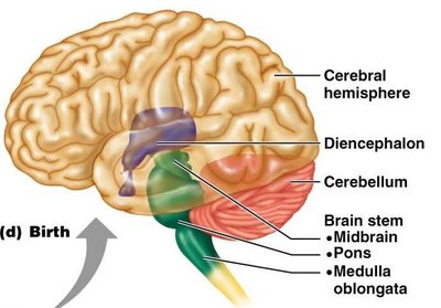
Cerebral hemispheres: Largest portion, responsible for higher functions.
Diencephalon: Includes thalamus, hypothalamus, and epithalamus.
Brain stem: Composed of midbrain, pons, and medulla oblongata.
Cerebellum: Coordinates movement and balance.
Distribution of Gray and White Matter
Structural Organization
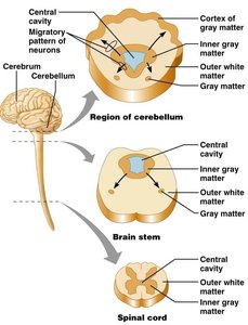
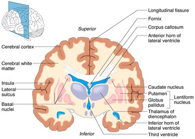
The brain and spinal cord are organized into gray and white matter, which differ in composition and function.
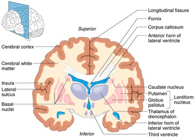
Gray matter: Contains neuron cell bodies and unmyelinated processes; forms the cortex in cerebrum and cerebellum.
White matter: Composed of myelinated fiber tracts; located deep in cerebrum and cerebellum, superficial in spinal cord.
Brain Ventricles
Fluid-Filled Spaces
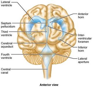
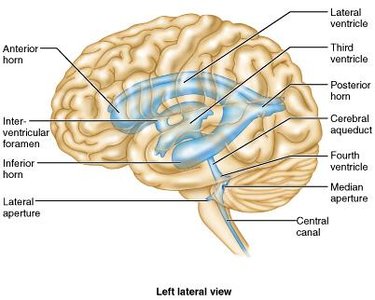
The brain contains interconnected ventricles filled with cerebrospinal fluid (CSF), which cushions and nourishes neural tissue.
Lateral ventricles: C-shaped chambers deep in cerebral hemispheres.
Third ventricle: Slit-like space between thalamic halves.
Fourth ventricle: Located between brain stem and cerebellum; connects to central canal of spinal cord.
Cerebral Hemispheres and Lobes
Surface Features and Divisions
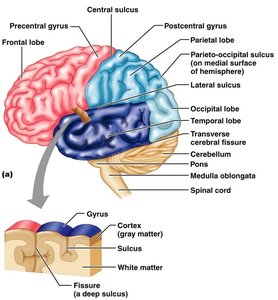
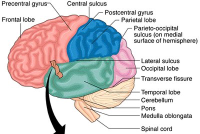
The cerebral hemispheres constitute about 80% of the brain's mass. The surface is marked by gyri (ridges), sulci (shallow grooves), and fissures (deep grooves).
Longitudinal fissure: Separates right and left hemispheres.
Transverse fissure: Separates cerebrum from cerebellum.
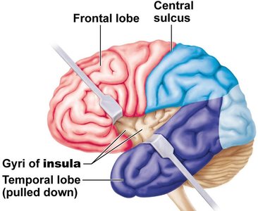
Lobes: Frontal, parietal, temporal, occipital, and insula (deep to other lobes).
Central sulcus: Separates frontal and parietal lobes.
Lateral sulcus: Separates frontal and temporal lobes.
Parieto-occipital sulcus: Separates parietal and occipital lobes.
Cerebral Cortex
Functional Organization
The cerebral cortex is the center of consciousness and higher brain functions. It is only 2–4 mm thick but accounts for about 40% of the brain's mass due to its extensive folding.
Contains: Neuron cell bodies, dendrites, unmyelinated axons, glial cells.
Three functional areas: Motor, sensory, and association.
Lateralization: Hemispheres are not equal in function; each controls the opposite side of the body.
Motor Areas of the Cerebral Cortex
Control of Voluntary Movement
Motor areas are located in the posterior part of the frontal lobes and are responsible for voluntary movement.
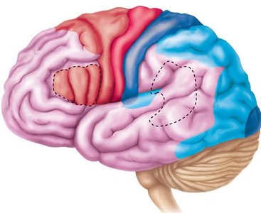
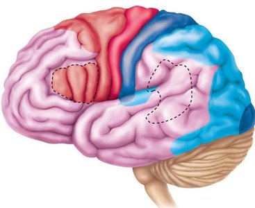
Primary motor cortex: Located in the precentral gyrus; controls precise, skilled movements.
Premotor cortex: Anterior to primary motor cortex; involved in learned, patterned movements.
Broca’s area: Motor center for speech; controls muscles of tongue, throat, lips.
Frontal eye field: Controls voluntary eye movements.
Motor Homunculus
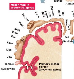
The motor homunculus is a map showing the regions of the precentral gyrus that control skeletal muscles of each body part. The size of each body part on the map reflects the number of neurons dedicated to its control.
Contralateral control: Each hemisphere controls the opposite side of the body.
Specialization: Large areas for hands, face, and tongue due to their complex movements.
Sensory Areas of the Cerebral Cortex
Somatosensory Cortex
The primary somatosensory area receives inputs from peripheral sensory receptors and localizes sensations. The somatosensory association area integrates and analyzes sensory information for object identification.
Spatial discrimination: Identifies the area of the body being stimulated.
Association area: Uses memories and experiences for identification without visual input.
Visual Cortex
Located in the occipital lobe, the visual cortex receives impulses from the eyes via the thalamus. It processes color, form, and movement.
Optic tract: Carries information from the visual field to the thalamus.
Lateral geniculate nucleus: Thalamic relay for visual information.
Auditory and Olfactory Cortex
The auditory cortex in the temporal lobe processes pitch, rhythm, and loudness, while the olfactory cortex in the piriform lobe provides conscious awareness of smells.
Auditory association area: Identifies sounds using memories.
Olfactory cortex: Processes smell information.
Association Areas and Brain Lateralization
Multimodal Association Areas
These areas integrate information from multiple senses and are involved in intellect, learning, recall, personality, pattern recognition, and emotional responses.
Anterior association area: Prefrontal cortex; involved in judgment, planning, and personality.
Posterior association area: Temporal, parietal, and occipital lobes; involved in pattern recognition and scene awareness.
Limbic association area: Emotional impact and sense of danger.
Brain Lateralization
Each hemisphere is dominant for certain activities. The left hemisphere is usually dominant for language, math, and logic, while the right is dominant for creative skills and visual-spatial abilities.
Hemispheric dominance: Most people are left-hemisphere dominant and right-handed.
Reversed dominance: Occurs in about 10% of people.
Cerebral White Matter and Basal Nuclei
White Matter Connections
Myelinated fibers in the CNS provide three types of connections:
Commissural fibers: Connect hemispheres (e.g., corpus callosum).
Association fibers: Connect neurons within one hemisphere.
Projection fibers: Connect cerebral hemispheres to other CNS parts.
Basal Nuclei
Basal nuclei are deep masses of gray matter involved in regulating slow, sustained motor movements and inhibiting unnecessary movements. Dysfunction is associated with Parkinson’s disease.
Diencephalon: Thalamus, Hypothalamus, and Epithalamus
Thalamus
The thalamus is the major relay station for sensory impulses, forming the lateral walls of the third ventricle. It processes sensory information and provides crude recognition of sensation.
Hypothalamus
The hypothalamus forms the bottom of the third ventricle and is connected to the pituitary gland. It regulates autonomic functions, emotional responses, body temperature, food and water intake, sleep-wake cycles, and endocrine functions.
Epithalamus
The epithalamus contains the pineal gland, which secretes melatonin and is involved in sleep-wake cycles. It is also a site for CSF production.
Brain Stem: Midbrain, Pons, and Medulla Oblongata
Structure and Function
The brain stem is composed of the midbrain, pons, and medulla oblongata. It is involved in automatic behaviors necessary for survival and provides pathways for communication between brain regions.
Midbrain: Connects pons to diencephalon; contains cerebral aqueduct.
Pons: Conduction pathways; origin of several cranial nerves.
Medulla oblongata: Maintains internal homeostasis; cardiovascular and respiratory centers.
Cerebellum
Structure and Function
The cerebellum is the second-largest brain region, separated from the cerebrum by the transverse fissure. It coordinates intended movements with actual movements, receiving sensory input from proprioceptors and equilibrium receptors.
Folia: Folds of the cerebellar cortex.
Arbor vitae: White matter tracts resembling a tree.
Purkinje neurons: Refine motor activities.
Functional Systems: Limbic System and Reticular Formation
Limbic System
The limbic system encircles the brain stem and is the emotional center of the brain. It is involved in emotional behavior, memory, and pleasure/pain responses.
Reticular Formation
The reticular formation is a network of gray matter nuclei in the brain stem, involved in sensory, integrative, and motor functions. The reticular activating system (RAS) maintains consciousness and filters repetitive stimuli.
Protection of the Brain
Protective Mechanisms
The brain is protected by several mechanisms:
Scalp hair: Prevents sunstroke.
Cranium: Bony protection.
Meninges: Three connective tissue membranes (dura mater, arachnoid, pia mater).
Cerebrospinal fluid (CSF): Cushions and nourishes the brain.
Blood-brain barrier: Separates CNS from bloodstream.
Meninges
Dura mater: Outermost, tough layer.
Arachnoid membrane: Middle, loose connective tissue; contains CSF.
Pia mater: Innermost, thin layer; follows brain contours.
Cerebrospinal Fluid (CSF)
CSF protects against injury, serves as a circulatory system, and maintains a constant chemical environment. It is produced by choroid plexuses and reabsorbed by arachnoid villi.
Blood-Brain Barrier
The blood-brain barrier regulates molecule penetration into the brain, protecting it from harmful substances. It is formed by tight junctions, basal lamina, and astrocytes.
Homeostatic Imbalances of the Brain
Traumatic Brain Injuries
Concussion: Temporary cognitive impairment.
Contusion: Bleeding and bruising.
Laceration: Tearing of brain tissue.
Hemorrhage: Bleeding increases intracranial pressure.
Cerebrovascular Accidents (Stroke)
Ischemia: Reduced blood flow causes tissue death.
Transient ischemic attack (TIA): Temporary reduction in blood flow.
Degenerative Brain Diseases
Alzheimer’s disease: Memory loss, cognitive deficits, abnormal protein deposits.
Parkinson’s disease: Degeneration of dopamine neurons, tremor, rigidity.
Cerebral palsy: Damage to motor areas, poor muscle control.
The Spinal Cord
Anatomy and Protection
The spinal cord is located within the vertebral column and protected by spinal meninges (dura mater, arachnoid, pia mater). The epidural space contains adipose and loose connective tissue.
External Anatomy
Cervical and lumbar enlargements: Nerves for upper and lower extremities.
Conus medullaris: Tapered end of the spinal cord.
Filum terminale: Pia mater extension attaching cord to coccyx.
Cauda equina: Bundle of spinal nerves at cord's end.
Cross-Sectional Anatomy
The spinal cord has an "H" shaped gray matter surrounded by white matter. The central canal contains CSF. Gray matter is divided into anterior (ventral), posterior (dorsal), and lateral horns.
PNS and Spinal Cord
Dorsal root: Afferent sensory nerves.
Posterior horn: Somatic and visceral sensory fibers.
Anterior horn: Motor neurons.
Ventral root: Efferent motor nerves.
Reflex Arcs
Reflex arcs are basic connections in the spinal cord, ranging from simple two-cell reflexes to complex circuits with interneurons.
White Matter of the Spinal Cord
White matter contains conduction tracts named by their origin and destination. Examples include fasciculi cuneatus and gracilis (fine touch), spinothalamic tracts (pain, temperature), and spinocerebellar tracts (proprioception).
Summary Table: Major Subdivisions of the Brain
Subdivision | Main Components | Function |
|---|---|---|
Cerebral Hemispheres | Frontal, parietal, temporal, occipital, insula | Higher functions, consciousness, voluntary movement |
Diencephalon | Thalamus, hypothalamus, epithalamus | Sensory relay, homeostasis, endocrine regulation |
Brain Stem | Midbrain, pons, medulla oblongata | Automatic behaviors, cranial nerve origin |
Cerebellum | Folia, arbor vitae, vermis | Coordination, balance, motor refinement |
Additional info: Academic context was added to clarify functions, structural relationships, and clinical relevance for exam preparation.
