 뒤로
Chapter 14: The Autonomic Nervous System – Structure and Function
스터디 가이드 - 스마트 노트
 자료에 맞춘 맞춤형 노트, 핵심 정의, 예시, 맥락을 확장해 제공합니다.
The Autonomic Nervous System (ANS)
Place of the ANS in the Structural Organization of the Nervous System
The Autonomic Nervous System (ANS) is a major motor division of the Peripheral Nervous System (PNS), responsible for involuntary control of internal organs, glands, and smooth and cardiac muscle. It is distinct from the somatic nervous system, which controls voluntary skeletal muscle movements.
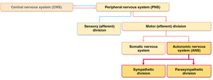
Motor Division of the PNS: ANS vs. Somatic Nervous System
The ANS operates through a two-neuron relay system, while the somatic nervous system uses a single motor neuron. The ANS regulates involuntary effectors such as cardiac muscle, smooth muscle, and glandular epithelial tissue.
ANS: Two-neuron relay (preganglionic and postganglionic neurons), ganglia outside CNS, effectors include cardiac muscle, smooth muscle, glands, neurotransmitters are acetylcholine (Ach) and norepinephrine (NE), effects can be excitatory or inhibitory.
Somatic Nervous System: Single motor neuron from CNS, effectors are skeletal muscle, neurotransmitter is Ach, effect is always excitatory.
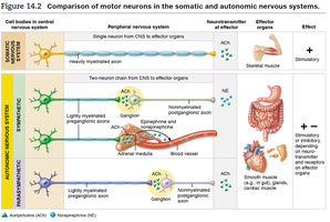
Two Divisions of the ANS: Sympathetic and Parasympathetic
The ANS is divided into two main branches: the Sympathetic Nervous System and the Parasympathetic Nervous System. Both divisions use a two-neuron relay and often innervate the same tissues, producing opposing effects (dual innervation).
Preganglionic neuron: Cell body within CNS, axon leaves CNS, uses Ach as neurotransmitter.
Postganglionic neuron: Synapses with effectors (cardiac muscle, smooth muscle, glands).
Dual Innervation: Both divisions innervate a tissue, producing opposite effects (e.g., sympathetic increases heart rate, parasympathetic decreases heart rate).
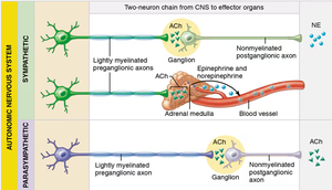
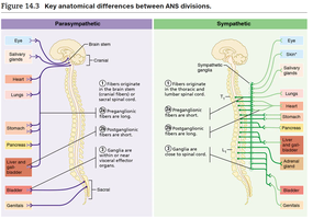
Overview of Differences: Sympathetic vs. Parasympathetic Division
The two divisions of the ANS differ in their anatomical origins, ganglia locations, fiber lengths, neurotransmitters, and physiological effects.
Sympathetic Division: Originates from thoracolumbar regions (T1-L2), ganglia close to spinal cord, short preganglionic fibers, long postganglionic fibers, widespread effects, neurotransmitters: Ach (preganglionic), NE (postganglionic), "Fight or Flight" response.
Parasympathetic Division: Originates from craniosacral regions (brainstem and S2-S4), ganglia close to or within effectors, long preganglionic fibers, short postganglionic fibers, localized effects, neurotransmitter: Ach (both pre- and postganglionic), "Rest and Digest" response.
Comparison of Effects of Innervation
The sympathetic and parasympathetic divisions produce distinct physiological effects on target organs.
Sympathetic: Pupillary dilation, increased heart rate and force, bronchiole dilation, increased sweat gland activity, decreased digestive and urinary activity, increased vasoconstriction and blood pressure, causes ejaculation/vaginal contraction.
Parasympathetic: Pupillary constriction, decreased heart rate and blood pressure, bronchiole constriction, increased salivary gland activity, increased digestive and urinary activity, causes erection.
Sympathetic Nervous System
Sympathetic Tone and Vasomotor Tone
Sympathetic tone refers to the baseline level of sympathetic activity, which is the main regulator of blood vessel diameter. Vasomotor tone is the state of partial constriction of blood vessels, allowing rapid adjustments in blood pressure.
If blood pressure drops, sympathetic activity increases, causing vasoconstriction and raising blood pressure.
If blood pressure is elevated, sympathetic activity decreases, causing vasodilation and lowering blood pressure.
Sympathetic Nervous System: Thoracolumbar Division
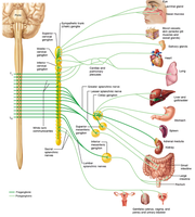
Preganglionic fibers arise from the thoracic and lumbar regions (T1-L2) of the spinal cord. These fibers synapse with ganglia (sympathetic chain ganglia, collateral ganglia, adrenal medullae), and postganglionic fibers are long (except for adrenal medullae).
Pathways of Innervation
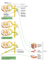
Sympathetic Chain Ganglia: Preganglionic fibers exit spinal cord and synapse at the same, upper, or lower level.
Collateral Ganglia: Preganglionic fibers form splanchnic nerves and synapse anterior to the spinal cord (e.g., celiac, superior mesenteric, inferior mesenteric ganglia).
Adrenal Medullae: Long preganglionic fibers synapse with specialized cells in the adrenal medullae, releasing NE and epinephrine.
Parasympathetic Nervous System
Craniosacral Division
Preganglionic fibers arise from the brainstem and sacral region (S2-S4) of the spinal cord. These fibers synapse with terminal ganglia close to or within target tissues. The cranial portion involves CN III, VII, IX, and X (vagus nerve supplies ~75% of output).
Neurotransmitters and Receptors of the ANS
Acetylcholine (Ach) and Cholinergic Receptors
Acetylcholine is released by all preganglionic fibers of the ANS, all parasympathetic postganglionic fibers, and a few sympathetic postganglionic fibers (targeting sweat glands). Cholinergic receptors on target cells bind Ach.
Nicotinic Receptors: Always excitatory; found on all postganglionic neurons and neuromuscular junctions.
Muscarinic Receptors: Can be excitatory or inhibitory; found on all parasympathetic targets, excitatory in most, inhibitory in cardiac muscle.
Norepinephrine (NE) and Adrenergic Receptors
Norepinephrine is released by most sympathetic postganglionic fibers. Adrenergic receptors on target cells bind NE.
Alpha Receptors: α1 (excitatory, e.g., vasoconstriction, pupil dilation), α2 (inhibitory).
Beta Receptors: β1 (excitatory, e.g., increased heart rate), β2 (mostly inhibitory, e.g., bronchiole dilation), β3 (excitatory, e.g., increased lipolysis in adipose tissue).
Visceral Reflexes
Structure and Function
Visceral reflexes are automatic responses to sensory information (chemical changes, stretch, temperature) detected by receptors. They involve the same elements as somatic reflexes: receptor, sensory neuron, integration center, motor neuron(s), and effector.
Short Reflexes: Bypass CNS, primarily in the enteric nervous system (controls gastrointestinal activity).
Long Reflexes: Use two motor neurons, coordinate multiple organ systems, more common than short reflexes.
Central Control of the ANS
Multiple Levels of Motor Control
The ANS is regulated by the CNS, with key centers including the medulla oblongata (processing complex visceral reflexes), hypothalamus (major integration center), thalamus, limbic system, and cerebral cortex. Higher brain centers influence ANS activity, integrating emotions, memory, and behavior.
Medulla Oblongata: Controls cardiac, vasomotor, swallowing, coughing, and respiratory centers.
Hypothalamus: Communicates with limbic system and cerebral cortex, influences medulla oblongata directly and indirectly.
Spinal Cord: T1–L2 (sympathetic reflexes), S2–S4 (parasympathetic reflexes).
