 뒤로
뒤로Chapter 14: The Brain and Cranial Nerves - Anatomy & Physiology Study Notes
스터디 가이드 - 스마트 노트
 자료에 맞춘 맞춤형 노트, 핵심 정의, 예시, 맥락을 확장해 제공합니다.
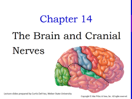
Chapter 14: The Brain and Cranial Nerves
Introduction to the Brain
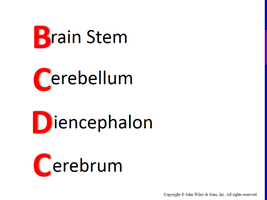
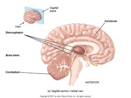
The brain is the central organ of the nervous system, responsible for processing sensory information, coordinating responses, and regulating behavior, intellect, emotions, and memory. It is divided into four main areas: the brain stem, cerebellum, diencephalon, and cerebrum.
Main Areas of the Brain
Brain Stem: Controls basic life functions and connects the brain to the spinal cord.
Cerebellum: Coordinates movement, balance, and posture.
Diencephalon: Contains the thalamus and hypothalamus, involved in sensory relay and homeostasis.
Cerebrum: Responsible for higher functions such as reasoning, memory, and voluntary movement.
Protective Coverings of the Brain
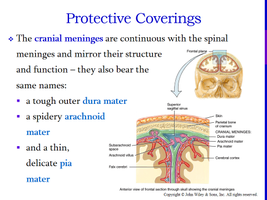
The brain is protected by cranial bones and three layers of meninges:
Dura mater: Tough, outer layer
Arachnoid mater: Spidery, middle layer
Pia mater: Thin, delicate inner layer
Extensions of the dura mater (falx cerebri, falx cerebelli, tentorium cerebelli) separate parts of the brain.
Brain Blood Flow and the Blood-Brain Barrier
The brain receives about 20% of the body's blood supply and oxygen. Blood is supplied by the internal carotid and vertebral arteries and drained by the jugular veins. The blood-brain barrier (BBB) protects brain tissue by restricting passage of harmful substances, formed by tight junctions between endothelial cells and astrocyte end-feet.
Clinical Connection: BBB breakdown can allow toxins or drugs to enter brain tissue, complicating treatment of CNS disorders.
Cerebrospinal Fluid (CSF)
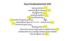
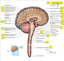
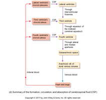
CSF is a clear fluid that cushions the brain and spinal cord, transports nutrients, and removes waste. It is produced by ependymal cells in the choroid plexuses of the ventricles and circulates through the brain and spinal cord.
Functions: Mechanical protection, chemical protection, circulation of nutrients and waste.
Production: Choroid plexuses in ventricles
Flow: Lateral ventricles → Interventricular foramina → Third ventricle → Cerebral aqueduct → Fourth ventricle → Median/lateral apertures → Subarachnoid space → Arachnoid villi → Dural venous sinuses
Reabsorption: Arachnoid villi into dural sinuses
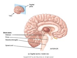
Brain Stem
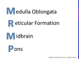
The brain stem is composed of four main parts: medulla oblongata, reticular formation, midbrain, and pons. It is essential for basic life functions and acts as a relay between the brain and spinal cord.
Medulla Oblongata
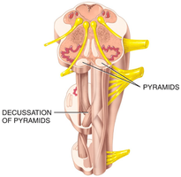
The medulla contains motor and sensory tracts, pyramids (where motor tracts decussate), and vital centers for cardiovascular and respiratory control. The inferior olivary nucleus relays proprioceptive signals to the cerebellum.
Pyramids: Motor tracts that cross over to control the opposite side of the body (decussation).
Vital Centers: Control heart rate, breathing, swallowing, vomiting, coughing, sneezing.
Inferior Olivary Nucleus: Relays impulses from proprioceptors to cerebellum.
Reticular Formation
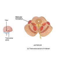
The reticular formation is a network of gray and white matter involved in sensory and motor functions. The reticular activating system (RAS) maintains consciousness and filters sensory information.
Midbrain
The midbrain contains cerebral peduncles (motor tracts), superior colliculi (visual reflexes), inferior colliculi (auditory reflexes), substantia nigra (dopamine release, muscle control), and red nucleus (voluntary limb movement).
Pons
The pons acts as a bridge connecting the spinal cord to the brain and helps control breathing through the pontine respiratory group.
Cerebellum
The cerebellum is responsible for coordinating movement, maintaining posture, equilibrium, and balance. It consists of the vermis (central area), hemispheres (lateral wings), cerebellar peduncles, and arbor vitae (white matter).
Diencephalon
The diencephalon includes the thalamus (sensory relay), hypothalamus (homeostasis, endocrine regulation), and epithalamus (pineal gland for melatonin, habenular nuclei for emotional response to odors). Circumventricular organs monitor blood chemistry and coordinate endocrine and nervous system activities.
Cerebrum
The cerebrum is the largest part of the brain, responsible for higher functions such as reasoning, memory, and voluntary movement. It consists of the cortex (gray matter), white matter tracts, basal nuclei (automatic movements, muscle tone), and limbic system (emotions, memory).
Lobes: Frontal (motor), Parietal (somatosensory), Occipital (visual), Temporal (auditory/olfactory), Insula (deep)
Corpus Callosum: Connects hemispheres
Hemispheric Lateralization: Left (language, reasoning), Right (artistic, spatial)
Functional Organization of the Cerebral Cortex
Sensory Areas: Reception and interpretation of sensory impulses (primary somatosensory, visual, auditory, gustatory, olfactory areas)
Motor Areas: Voluntary muscle movement (primary motor area, Broca's area for speech)
Association Areas: Complex functions (memory, emotions, reasoning, intelligence)
Brain Waves
Electrical potentials generated by brain cells are called brain waves, recorded as an electroencephalogram (EEG). EEGs are used to diagnose neurological disorders and confirm brain death.
Cranial Nerves
There are twelve pairs of cranial nerves, each with specific sensory, motor, or mixed functions. They originate from the brain and are essential for sensory perception and motor control.
Nerve | Function | Type |
|---|---|---|
I Olfactory | Smell | Sensory |
II Optic | Vision | Sensory |
III Oculomotor | Eye movements, pupil size | Motor |
IV Trochlear | Eye movements | Motor |
V Trigeminal | Chewing, sensations of head & face | Both |
VI Abducens | Abduction of eye | Motor |
VII Facial | Facial expressions, taste | Both |
VIII Vestibulocochlear | Balance, hearing | Sensory |
IX Glossopharyngeal | Swallowing, taste, blood pressure | Both |
X Vagus | Sensations and movement of organs | Both |
XI Accessory | Shoulder, head movements | Motor |
XII Hypoglossal | Tongue movements | Motor |
Aging and the Nervous System
Age-related changes include loss of neurons, decreased nerve impulse conduction, slower voluntary movements, increased reflex time, and altered sensory functions (vision, hearing, taste, smell, touch).
Summary Table: Principal Parts and Functions of the Brain
Brain Area | Main Function |
|---|---|
Brain Stem | Basic life functions, relay between brain and spinal cord |
Cerebellum | Coordination, balance, posture |
Diencephalon | Sensory relay, homeostasis, endocrine regulation |
Cerebrum | Higher functions: reasoning, memory, voluntary movement |
