 뒤로
Chapter 21: The Immune System – Innate and Adaptive Body Defenses
스터디 가이드 - 스마트 노트
 자료에 맞춘 맞춤형 노트, 핵심 정의, 예시, 맥락을 확장해 제공합니다.
The Immune System: Overview
Introduction
The immune system is a functional system that provides resistance to disease-causing microorganisms such as bacteria, fungi, and viruses. It consists of a diverse array of molecules and immune cells, rather than a single anatomical organ. The body has three lines of defense: innate (nonspecific) defenses and adaptive (specific) defenses, which together provide immunity. 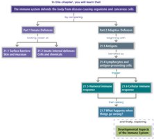
Organization of Defenses
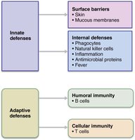
Innate Defenses: Surface barriers (skin, mucous membranes) and internal defenses (phagocytes, NK cells, inflammation, antimicrobial proteins, fever).
Adaptive Defenses: Humoral immunity (B cells) and cellular immunity (T cells).
Part 1 – Innate Defenses
21.1 First Line: Surface Barriers
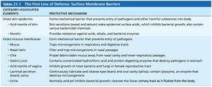
Surface barriers, including skin and mucous membranes, act as physical and chemical barriers to most microorganisms. Keratin in the skin is resistant to acids, bases, bacterial enzymes, and toxins. These barriers also produce protective chemicals such as acidic secretions, enzymes, and mucin.
Acid Mantle: Acidity of skin, vaginal, and stomach secretions inhibits bacterial growth.
Enzymes: Lysozyme in saliva, mucus, and tears kills bacteria; stomach enzymes destroy microorganisms.
Mucin: Sticky mucus traps microorganisms in digestive and respiratory tracts.
Respiratory Modifications: Mucus-coated hairs in the nose trap particles; cilia sweep mucus toward the mouth.
21.2 Second Line: Innate Internal Defenses
When surface barriers are breached, internal defenses are activated. These include phagocytes, natural killer (NK) cells, inflammation, antimicrobial proteins, and fever. Many cells have pattern recognition receptors to identify pathogens.
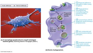
Phagocytes: White blood cells that engulf and digest invaders. Neutrophils are the most abundant, while macrophages are chief phagocytic cells (free or fixed in tissues).
Phagocytosis: Phagocytes adhere to pathogens, engulf them in a phagosome, which fuses with a lysosome to form a phagolysosome. Lysosomal enzymes digest the pathogen, and waste is exocytosed.
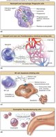
Natural Killer (NK) Cells: Kill cancer and virus-infected cells by inducing apoptosis. They attack cells lacking "self" MHC proteins and secrete chemicals to enhance inflammation.
Inflammation: A nonspecific response to injury, characterized by redness, heat, swelling, and pain. It prevents spread of damage, disposes of debris, alerts the adaptive system, and sets the stage for repair.
Stages of Inflammation
Inflammatory chemical release (e.g., histamine, cytokines)
Vasodilation and increased vascular permeability (causes hyperemia and edema)
Phagocyte mobilization (leukocytosis, margination, diapedesis, chemotaxis)
Clinical View: Inflammation Aftermath
Pus: Mixture of dead neutrophils, tissue, and pathogens.
Abscess: Sac of pus walled off by collagen fibers.
Ulcer: Open sore from loss of superficial tissue.
Granulomas: Tumor-like growths from resistant bacteria.
Antimicrobial Proteins
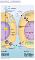
Interferons (IFN): Proteins secreted by virus-infected cells to warn neighbors and block viral reproduction. Also activate NK cells and macrophages.
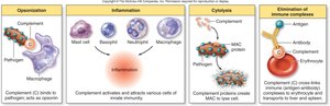
Complement: Group of ~20 blood proteins that enhance both innate and adaptive defenses. Causes cell lysis, opsonization, and amplifies inflammation.
Fever
Definition: Abnormally high body temperature in response to infection.
Mechanism: Pyrogens released by leukocytes and macrophages act on hypothalamus.
Benefits: Enhances immune response, increases metabolic rate, suppresses bacterial growth.
Risks: Sustained high fever can be harmful.
Part 2 – Adaptive Defenses
Characteristics of Adaptive Immunity
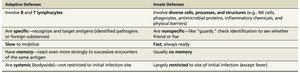
The adaptive immune system is specific, systemic, and has memory. It involves lymphocytes (B and T cells) and takes longer to mobilize than innate defenses.
Humoral Immunity: Antibodies produced by B cells target extracellular pathogens.
Cellular Immunity: T cells target infected, cancerous, or foreign cells.
21.3 Antigens
Definition: Substances that provoke an immune response; usually large, foreign molecules.
Self-antigens: Proteins on cell surfaces not antigenic to self but may be to others (e.g., MHC proteins).
21.4 Lymphocytes and Antigen-Presenting Cells (APCs)
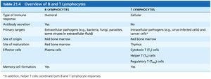
B Lymphocytes: Oversee humoral immunity.
T Lymphocytes: Oversee cellular immunity.
APCs: Engulf antigens and present fragments to T cells. Dendritic cells, macrophages, and B cells are key APCs.
Lymphocyte Development, Maturation, and Activation
Origin: Both B and T cells originate in red bone marrow.
Maturation: T cells mature in thymus; B cells in bone marrow. Develop immunocompetence and self-tolerance.
Seeding: Naive lymphocytes colonize secondary lymphoid organs.
Antigen Encounter: Activation occurs when antigen binds to receptor.
Proliferation and Differentiation: Activated lymphocytes form clones; most become effector cells, some become memory cells.
Antigen-Presenting Cells (APCs)
Dendritic Cells: Phagocytize pathogens and present antigens to T cells.
Macrophages: Present antigens and activate themselves.
B Lymphocytes: Present antigens to helper T cells.
Humoral Immune Response
Activation and Differentiation of B Cells
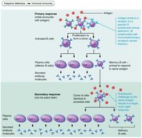
B cells are activated by antigen binding (clonal selection).
Most clones become plasma cells, secreting antibodies; others become memory cells.
Immunological Memory
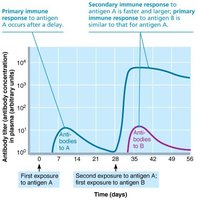
Primary Response: Lag period of 3–6 days; antibody levels peak in 10 days.
Secondary Response: Faster, stronger, longer-lasting due to memory cells.
Active and Passive Humoral Immunity
Active Immunity: B cells produce antibodies after exposure (natural or artificial via vaccine).
Passive Immunity: Antibodies are introduced (e.g., maternal antibodies, antivenom); no memory formed.
Antibodies (Immunoglobulins)
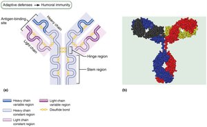
Y-shaped proteins secreted by plasma cells; bind specifically to antigens.
Variable regions form antigen-binding sites; constant regions determine class and function.
Antibody Classes
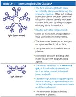
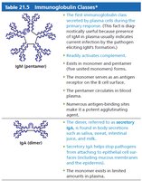
IgM: First released, pentamer, potent agglutinating agent.
IgA: Found in secretions, prevents pathogen entry.
IgD: B cell receptor.
IgG: Most abundant, crosses placenta, main secondary response antibody.
IgE: Active in allergies and parasitic infections.
Antibody Functions
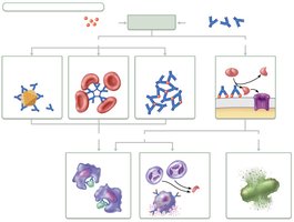
Neutralization: Block sites on pathogens/toxins.
Agglutination: Bind multiple antigens, forming clumps.
Precipitation: Cross-link soluble molecules.
Complement Activation: Leads to cell lysis, opsonization, and inflammation.
Cellular Immune Response
T Cells and Antigen Presentation
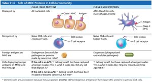
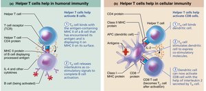
T cells respond only to processed antigen fragments presented by MHC proteins.
Class I MHC: Found on all cells except RBCs; present endogenous antigens to CD8 cells (cytotoxic T cells).
Class II MHC: Found on APCs; present exogenous antigens to CD4 cells (helper T cells).
Activation and Differentiation of T Cells
T cell receptors (TCRs) bind to antigen-MHC complex.
Activated T cells proliferate and differentiate into effector and memory cells.
Cytokines mediate cell development and responses.
Roles of Specific Effector T Cells
Helper T (TH) Cells: Activate B cells, other T cells, and macrophages; central to adaptive response.
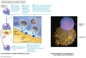
Cytotoxic T (TC) Cells: Directly attack and kill infected, cancerous, or foreign cells by inducing apoptosis.
Regulatory T (TReg) Cells: Suppress immune response, prevent autoimmunity.
Immune System Disorders
Immunodeficiency
SCID: Congenital deficit in B and T cells; fatal if untreated.
AIDS: HIV destroys helper T cells, leading to opportunistic infections.
Autoimmune Diseases
Immune system attacks self tissues (e.g., rheumatoid arthritis, type 1 diabetes).
Caused by failure of self-tolerance, cross-reactivity, or new self-antigens.
Treatments include immunosuppressive drugs and cytokine blockers.
Hypersensitivities
Immediate (Type I): Allergies; IgE-mediated, rapid onset.
Subacute (Type II/III): IgM/IgG-mediated, slower onset.
Delayed (Type IV): T cell-mediated, appears in 1–3 days (e.g., contact dermatitis).
Developmental Aspects of the Immune System
Development and Influences
Stem cells develop in liver and spleen early, then bone marrow.
Lymphocyte development continues in bone marrow and thymus.
Immune function is influenced by nervous system and diet (vitamin D).
With age, immune function declines, increasing susceptibility to disease.
