 뒤로
Chapter 23: The Digestive System – Anatomy & Physiology Study Notes
스터디 가이드 - 스마트 노트
 자료에 맞춘 맞춤형 노트, 핵심 정의, 예시, 맥락을 확장해 제공합니다.
The Digestive System: Overview
Introduction
The digestive system is essential for converting food into nutrients that fuel and build the body's cells. It consists of a continuous muscular tube called the alimentary canal (gastrointestinal tract) and accessory organs that aid digestion. Understanding its structure and function is crucial for assessing digestive health and disorders.
Main Components
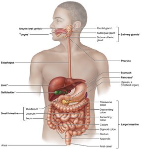
Alimentary Canal (GI Tract): Mouth, pharynx, esophagus, stomach, small intestine, large intestine, anus.
Accessory Organs: Teeth, tongue, gallbladder, salivary glands, liver, pancreas.
Major Digestive Processes
Six Essential Activities
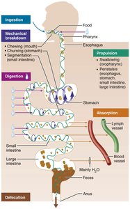
Ingestion: Intake of food.
Propulsion: Movement of food through the GI tract, including swallowing and peristalsis.
Mechanical Breakdown: Physical breakdown of food (chewing, churning, segmentation).
Digestion: Chemical breakdown of food by enzymes.
Absorption: Passage of nutrients into blood or lymph.
Defecation: Elimination of indigestible residues as feces.
Propulsion: Peristalsis vs. Segmentation
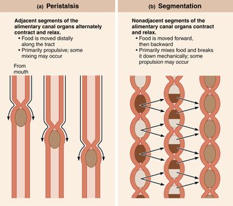
Peristalsis: Alternating waves of contraction and relaxation move food distally.
Segmentation: Local contractions mix food with digestive fluids, enhancing breakdown.
Structural Organization of the GI Tract
Layers of the GI Tract
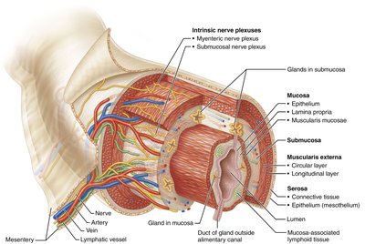
The walls of the alimentary canal have four basic layers:
Mucosa: Innermost layer; secretes mucus, enzymes, hormones; absorbs nutrients; protects against pathogens.
Submucosa: Areolar connective tissue; contains blood vessels, lymphatics, nerves.
Muscularis Externa: Smooth muscle; responsible for peristalsis and segmentation.
Serosa: Outermost layer; visceral peritoneum.
Peritoneum and Mesenteries
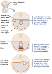
Peritoneum: Serous membrane lining the abdominopelvic cavity.
Visceral Peritoneum: Covers external surfaces of digestive organs.
Parietal Peritoneum: Lines the body wall.
Mesentery: Double layer of peritoneum; anchors organs, stores fat, provides routes for vessels and nerves.
Intraperitoneal Organs: Surrounded by peritoneum.
Retroperitoneal Organs: Located posterior to peritoneum.
Regulation of Digestive Activity
Enteric Nervous System
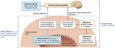
The GI tract has its own nervous system, the enteric nervous system (gut brain), which regulates digestive activities independently and in coordination with the central nervous system.
Submucosal Nerve Plexus: Controls glandular secretion and local blood flow.
Myenteric Nerve Plexus: Controls GI tract motility.
Short Reflexes: Mediated by enteric plexuses in response to internal stimuli.
Long Reflexes: Involve CNS and autonomic nerves, responding to internal and external stimuli.
Functional Anatomy: The Mouth
Oral Cavity Structure
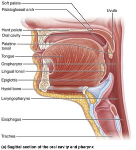
Boundaries: Lips, cheeks, palate, tongue.
Epithelium: Stratified squamous, keratinized in areas of friction.
Lips and Cheeks: Orbicularis oris and buccinator muscles.
Palate: Hard (bony) and soft (muscular) palate; uvula projects from soft palate.
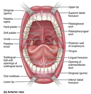
The Tongue
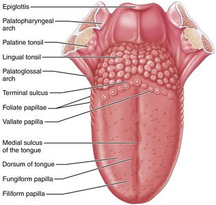
Muscle Bundles: Intrinsic (shape) and extrinsic (position) muscles.
Functions: Mixes food, forms bolus, initiates swallowing, speech, taste.
Papillae: Filiform (friction), fungiform (taste), vallate (taste), foliate (taste).
Lingual Frenulum: Attaches tongue to floor of mouth.
Salivary Glands
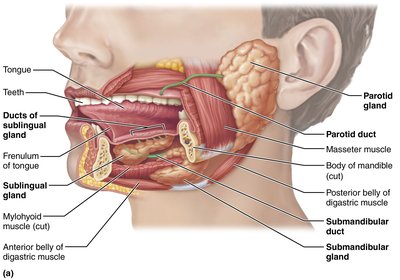
Major Glands: Parotid, submandibular, sublingual.
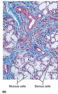
Secretory Cells: Serous (watery, enzyme-rich) and mucous (mucus).
Saliva: Cleanses mouth, moistens food, begins starch digestion (amylase), contains IgA, lysozyme, defensins.
Teeth
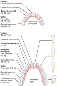
Types: Incisors (cutting), canines (tearing), premolars and molars (grinding).
Dentition: Deciduous (20) and permanent (32) teeth.
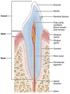
Structure: Crown (enamel), neck, root (cementum, periodontal ligament, dentin, pulp cavity).
Pharynx and Esophagus
Pharynx
Passageway: Oropharynx and laryngopharynx allow passage of food, fluids, air.
Muscle Layers: Skeletal muscle for swallowing.
Esophagus
Structure: Muscular tube; collapsed when not propelling food.
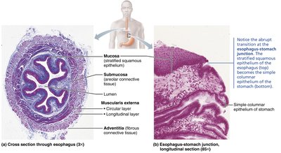
Layers: Mucosa (stratified squamous), submucosa (mucus glands), muscularis externa (skeletal/smooth muscle), adventitia.
Swallowing (Deglutition)
Buccal Phase: Voluntary, in mouth.
Pharyngeal-Esophageal Phase: Involuntary, via vagus nerve.
Summary Table: Digestive System Layers
Layer | Main Function | Key Features |
|---|---|---|
Mucosa | Secretion, absorption, protection | Epithelium, lamina propria, muscularis mucosae |
Submucosa | Support, vascular supply | Areolar connective tissue, blood/lymph vessels, nerves |
Muscularis Externa | Motility (peristalsis, segmentation) | Inner circular, outer longitudinal muscle layers |
Serosa | Protection, anchoring | Visceral peritoneum, areolar tissue |
Key Terms and Concepts
Peristalsis: Involuntary waves of contraction moving food through GI tract.
Segmentation: Local contractions mixing food with digestive fluids.
Mesentery: Double layer of peritoneum anchoring organs.
Enteric Nervous System: Intrinsic neural network regulating GI function.
Accessory Organs: Organs aiding digestion but not part of the GI tract.
Equations and Formulas
Alkaline Tide:
Dental Formula (Permanent):
Clinical Applications
Peritonitis: Inflammation of peritoneum, often from ruptured appendix.
Xerostomia: Dry mouth due to reduced saliva production.
Ankyloglossia: Restricted tongue movement from short lingual frenulum.
Dental Caries: Tooth decay from bacterial action.
Heartburn: Acid reflux into esophagus.
Summary
The digestive system is a complex network of organs and tissues that work together to ingest, break down, absorb, and eliminate food. Its structural and functional organization is essential for maintaining health and supporting metabolic needs. Understanding its anatomy and physiology provides a foundation for diagnosing and treating digestive disorders.
