 뒤로
Chapter 7: The Muscular System – Essentials of Anatomy & Physiology Study Notes
스터디 가이드 - 스마트 노트
 자료에 맞춘 맞춤형 노트, 핵심 정의, 예시, 맥락을 확장해 제공합니다.
The Muscular System
Introduction to Muscle Tissue
Muscle tissue is one of the four primary tissue types in the human body, specialized for contraction and movement. There are three types of muscle tissue: skeletal muscle, cardiac muscle, and smooth muscle. Each type has unique structural and functional characteristics.
Skeletal muscle: Voluntary, striated, attached to bones.
Cardiac muscle: Involuntary, striated, found only in the heart.
Smooth muscle: Involuntary, non-striated, found in walls of organs.
Skeletal Muscle Functions
Skeletal muscles perform five primary functions essential for movement and homeostasis:
Movement: Muscles pull on tendons to move bones.
Posture: Maintain body position and stabilize joints.
Support: Support soft tissues, especially in the abdominal and pelvic regions.
Guard entrances/exits: Encircle openings of digestive and urinary tracts.
Temperature regulation: Muscle contractions generate heat.
Organization of Skeletal Muscle Tissue
Structural Hierarchy
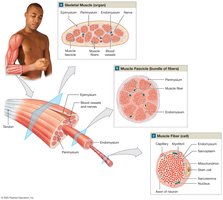
Skeletal muscle is organized into layers of connective tissue, blood vessels, nerves, and muscle fibers. Each muscle fiber is a single cell, and muscles are composed of bundles of these fibers.
Epimysium: Surrounds the entire muscle.
Perimysium: Divides muscle into fascicles (bundles of fibers).
Endomysium: Covers each muscle fiber, contains capillaries and nerves.
Connective Tissue Attachments
Collagen fibers from all three connective tissue layers merge to form:
Tendon: Attaches muscle to bone (bundle of fibers).
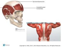
Aponeurosis: Broad sheet connecting muscles to each other.
Blood Vessels and Nerves
Skeletal muscles have an extensive blood supply to meet high energy demands. They are under voluntary control, requiring stimulation by the central nervous system. Axons innervate individual muscle fibers.
Features of Skeletal Muscle Fibers
Muscle Fiber Structure
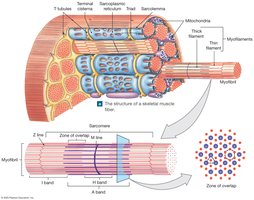
Skeletal muscle fibers are long, multinucleate cells with a striated appearance due to the arrangement of myofilaments.
Sarcolemma: Plasma membrane of the muscle cell.
Sarcoplasm: Cytoplasm of the muscle cell.
Transverse (T) tubules: Allow electrical impulses to reach the cell's interior.
Myofibrils and Myofilaments
Myofibrils are cylindrical structures composed of bundles of thick (myosin) and thin (actin) filaments. Their shortening causes muscle contraction.
Sarcoplasmic Reticulum and Triad
The sarcoplasmic reticulum (SR) is a specialized smooth endoplasmic reticulum that stores calcium ions. The triad consists of two terminal cisternae and one T tubule.
Sarcomere Structure and Function
Sarcomere Organization
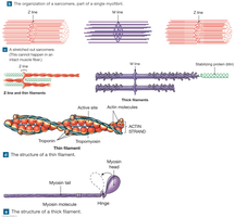
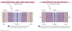
The sarcomere is the smallest functional unit of a muscle fiber, responsible for contraction. Its arrangement of thick and thin filaments produces the striated appearance.
Z lines: Boundaries of the sarcomere.
M line: Center of the sarcomere.
A band: Dark region with thick filaments.
I band: Light region with thin filaments.
H band: Region with only thick filaments.
Sliding Filament Theory
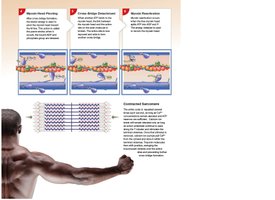
This theory explains how muscle contraction occurs. During contraction, thin filaments slide toward the center of the sarcomere, while thick filaments remain stationary. This shortens the sarcomere and produces tension.
Neuromuscular Junction and Muscle Contraction
Neuromuscular Junction (NMJ)
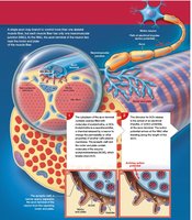
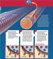
The NMJ is the site where a motor neuron communicates with a muscle fiber. Acetylcholine (ACh) is released from the neuron, binds to receptors on the muscle, and triggers an action potential.
The Contraction Cycle
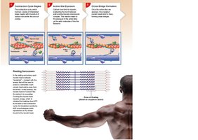
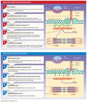
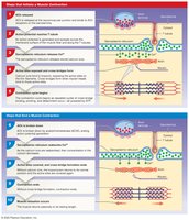
Muscle contraction begins when an action potential travels along the sarcolemma and T tubules, causing calcium release from the SR. Calcium binds to troponin, exposing actin's active sites for myosin binding.
Steps of Muscle Contraction and Relaxation
The process involves several steps, from ACh release to calcium reuptake and covering of actin's active sites.
Muscle Tension and Contraction Types
Muscle Twitch and Tension Development
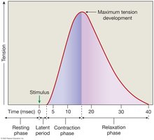
A muscle twitch is a single stimulus-contraction-relaxation cycle. Tension depends on the number of cross-bridges formed, fiber length, and stimulation frequency.
Latent period: Time between stimulus and contraction.
Contraction phase: Cross-bridge formation and tension increase.
Relaxation phase: Calcium reuptake and tension decrease.
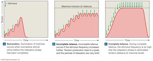
Summation and Tetanus
Summation is the addition of twitches for stronger contraction. Tetanus occurs when stimulation is so rapid that relaxation does not occur, resulting in maximum tension.
Motor Units and Recruitment
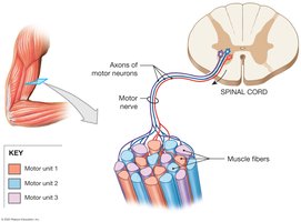
A motor unit consists of a motor neuron and all the muscle fibers it controls. Recruitment is the activation of more motor units to increase tension.
Muscle Tone and Atrophy
Muscle tone is the resting tension in muscles, important for posture. Atrophy is the loss of muscle mass due to inactivity or nerve damage.
Isotonic and Isometric Contractions
Isotonic: Muscle changes length (e.g., lifting a book).
Isometric: Muscle length remains the same (e.g., pushing against a wall).
Muscle Metabolism and Energy
ATP and Creatine Phosphate
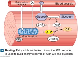
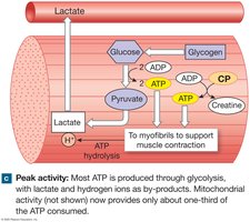
Muscle contraction requires ATP. At rest, excess ATP is stored as creatine phosphate (CP), which can quickly regenerate ATP during contraction.
Aerobic and Anaerobic Metabolism
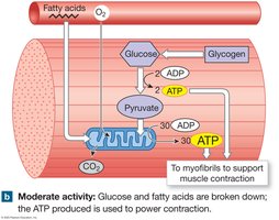
Aerobic metabolism: Occurs in mitochondria, requires oxygen, produces most ATP.
Glycolysis: Anaerobic, occurs in cytoplasm, produces ATP and lactic acid.
Muscle Fatigue and Recovery
Fatigue occurs when energy reserves are depleted or pH drops due to lactic acid. Recovery involves restoring oxygen, removing lactic acid, and replenishing energy stores.
Muscle Performance and Fiber Types
Fast vs. Slow Muscle Fibers
Fast fibers: Quick, powerful contractions, fatigue rapidly.
Slow fibers: Slower, sustained contractions, fatigue resistant.
Muscle conditioning can increase anaerobic or aerobic endurance.
Cardiac and Smooth Muscle Tissue
Cardiac Muscle
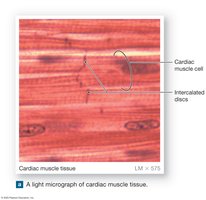
Cardiac muscle is found only in the heart, with cells connected by intercalated discs for synchronized contraction. It contracts automatically and relies on aerobic metabolism.
Smooth Muscle
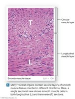
Smooth muscle is found in organ walls, lacks striations, and contracts over a wide range of lengths. It is involuntary and responds to hormones or neural input.
Major Muscles of the Body
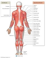
Axial and Appendicular Muscles
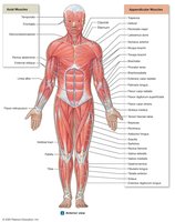
Axial muscles arise from the axial skeleton and position the head, spine, and rib cage. Appendicular muscles stabilize or move the limbs.
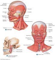
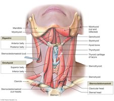
Muscles of the Head and Neck
These muscles control facial expressions, chewing, and movements of the neck.
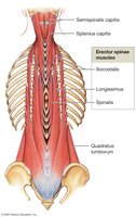
Muscles of the Spine
Spinal muscles maintain posture and allow movement of the head and spine.
Axial Muscles of the Trunk
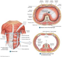
These include intercostals, diaphragm, obliques, and rectus abdominis, which are involved in breathing and trunk movement.
Muscles of the Pelvic Floor
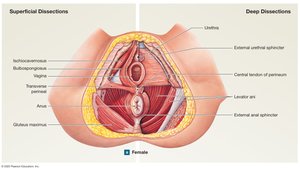
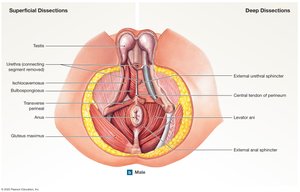
Pelvic floor muscles support pelvic organs and control movement through the anus and urethra.
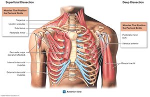
Muscles That Position the Pectoral Girdle
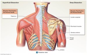
These muscles move and stabilize the shoulder.
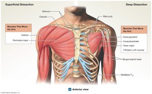
Muscles That Move the Arm
Include deltoid, rotator cuff muscles, pectoralis major, and latissimus dorsi.
Muscles That Move Forearm, Wrist, and Hand
Biceps brachii, triceps brachii, brachialis, and forearm flexors/extensors control elbow, wrist, and finger movements.
Muscles of the Pelvis and Lower Limbs
Gluteal muscles, adductors, quadriceps, hamstrings, and calf muscles move the hip, knee, ankle, and toes.
Effects of Aging and Exercise
Aging Effects
Muscle fibers decrease in diameter and elasticity.
Exercise tolerance and thermoregulation decline.
Recovery from injury is slower.
Exercise and Body Systems
Exercise increases oxygen consumption, heart rate, and respiration. The nervous and endocrine systems regulate these responses for homeostasis.
Comparison of Muscle Tissue Types
Table: Skeletal, Cardiac, and Smooth Muscle Comparison
Feature | Skeletal Muscle | Cardiac Muscle | Smooth Muscle |
|---|---|---|---|
Control | Voluntary | Involuntary | Involuntary |
Striations | Present | Present | Absent |
Location | Attached to bones | Heart | Walls of organs |
Cell shape | Long, cylindrical | Branched | Spindle-shaped |
Nucleus | Multinucleate | Single (usually) | Single |
Special features | Motor units, rapid contraction | Intercalated discs, automaticity | Can contract over wide range |
Additional info: This table summarizes the main differences between muscle tissue types, aiding in classification and understanding their functions.
