 뒤로
뒤로Chapter 7: The Skeleton – Axial Skeleton and Skull Anatomy
스터디 가이드 - 스마트 노트
 자료에 맞춘 맞춤형 노트, 핵심 정의, 예시, 맥락을 확장해 제공합니다.
The Skeletal System
Overview and Divisions
The skeletal system is a structural framework for the human body, composed of bones, cartilages, joints, and ligaments. It accounts for approximately 20% of body mass and is divided into two major divisions: the axial skeleton and the appendicular skeleton.
Axial skeleton: Consists of 80 bones in three regions: skull, vertebral column, and thoracic cage.
Appendicular skeleton: Includes bones of the limbs and girdles that attach them to the axial skeleton.
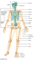
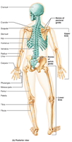
Axial Skeleton
Functions and Regions
The axial skeleton forms the longitudinal axis of the body, supports the head, neck, and trunk, and protects the brain, spinal cord, and thoracic organs. Its three major regions are:
Skull
Vertebral column
Thoracic cage
The Skull
General Structure
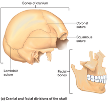
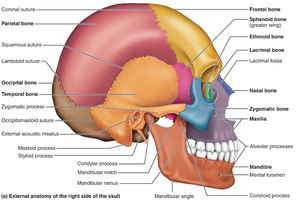
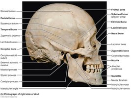
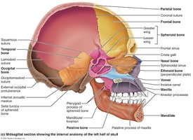
The skull is the most complex bony structure in the body, formed by two sets of bones: cranial bones and facial bones. Most skull bones are flat and firmly locked together by joints called sutures, which have a serrated, saw-tooth appearance.
Cranial bones (cranium): Enclose the brain and provide attachment sites for head and neck muscles.
Facial bones: Form the framework of the face, contain cavities for special sense organs, provide openings for air and food passage, secure teeth, and anchor facial muscles.
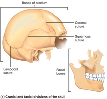
Skull Geography
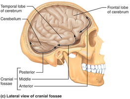
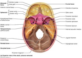
The cranium is divided into a vault (calvaria) and a base. The cranial vault forms the superior, lateral, and posterior portions of the skull, while the cranial base forms the inferior aspect. Internally, the base is divided into three cranial fossae: anterior, middle, and posterior, which house the brain.
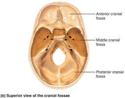
Skull Cavities and Openings
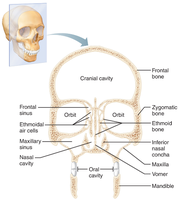
The skull contains several cavities, including the cranial cavity, middle and internal ear cavities, nasal cavity, and orbits. There are 85 named openings (foramina, canals, fissures) that provide passageways for the spinal cord, blood vessels, and cranial nerves.
Cranial Bones
Frontal Bone
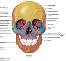
The frontal bone forms the anterior portion of the cranium, including the forehead (squamous region), superior wall of the orbits, and most of the anterior cranial fossa. Key features include the supraorbital margin, supraorbital foramen, glabella, and frontal sinuses.
Parietal Bones and Major Sutures
Two parietal bones form most of the superior and lateral aspects of the cranial vault. Four major sutures mark their articulations:
Coronal suture: Parietal and frontal bones
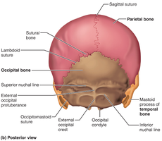
Sagittal suture: Right and left parietal bones
Lambdoid suture: Parietal and occipital bones
Squamous suture: Parietal and temporal bones
Occipital Bone
The occipital bone forms the posterior wall and posterior cranial fossa. It articulates with parietal, temporal, and sphenoid bones. Key features include:
Foramen magnum: Passage for the spinal cord
Occipital condyles: Articulate with the first vertebra
External occipital protuberance and crest: Sites for ligament and muscle attachment
Superior and inferior nuchal lines: Muscle attachment sites
Temporal Bones
Paired temporal bones make up the inferolateral aspects of the skull and parts of the cranial base. They have three major regions:
Squamous: Zygomatic process forms zygomatic arch; mandibular fossa forms part of temporomandibular joint
Tympanic: Surrounds external acoustic meatus
Petrous: Houses middle and internal ear cavities; contains several foramina for nerves and arteries
Sphenoid Bone
The sphenoid bone is a complex, bat-shaped bone that articulates with all other cranial bones. It contains sphenoidal sinuses and the sella turcica, which houses the pituitary gland. The bone has three pairs of processes (greater wings, lesser wings, pterygoid processes) and several foramina for nerves and arteries.
Ethmoid Bone
The ethmoid bone is the deepest skull bone, forming the roof of the nasal cavity and part of the anterior cranial fossa. Key features include the cribriform plates, crista galli, perpendicular plate, ethmoidal air cells, and superior and middle nasal conchae.
Sutural Bones
Sutural bones are tiny, irregularly shaped bones that appear within sutures. Their significance is unknown, and not everyone has them.
Facial Bones
Overview
The facial skeleton consists of 14 bones, 12 of which are paired. The mandible and vomer are single bones. The facial bones include:
Mandible
Maxillary bones (2)
Zygomatic bones (2)
Nasal bones (2)
Lacrimal bones (2)
Palatine bones (2)
Vomer
Inferior nasal conchae (2)
Mandible
The mandible is the largest and strongest bone of the face, forming the lower jaw. It consists of a body (chin) and two upright rami. Key features include the mandibular angle, coronoid process, condylar process, alveolar process, mandibular symphysis, and foramina for nerves and blood vessels.
Maxillary Bones
The maxillary bones are medially fused to form the upper jaw and central facial skeleton. They hold the upper teeth, form the anterior nasal spine, and contribute to the hard palate, nose, and orbits. Maxillary sinuses flank the nasal cavity laterally.
Zygomatic Bones
Zygomatic bones form the cheekbones and inferolateral margins of the orbits. They articulate with the zygomatic processes of the temporal, frontal, and maxillary bones.
Nasal and Lacrimal Bones
Nasal bones form the bridge of the nose and attach to cartilage that forms the tip. Lacrimal bones form the medial walls of the orbits and contain the lacrimal fossa for tear drainage.
Palatine Bones and Vomer
Palatine bones are L-shaped, forming the posterior part of the hard palate and part of the nasal cavity and orbits. The vomer is a plow-shaped bone forming part of the nasal septum.
Inferior Nasal Conchae
Paired bones forming part of the lateral walls of the nasal cavity; they are the largest of three pairs of conchae.
The Hyoid Bone
Structure and Function
The hyoid bone is unique in that it does not articulate directly with any other bone. Located in the anterior neck, it is anchored by ligaments and serves as a movable base for the tongue and attachment site for muscles involved in swallowing and speech.
Special Characteristics of the Orbits and Nasal Cavity
Orbits
The orbits are cavities that encase the eyes and lacrimal glands, providing attachment sites for eye muscles. They are formed by parts of seven bones: frontal, sphenoid, zygomatic, maxilla, palatine, lacrimal, and ethmoid.
Nasal Cavity
The nasal cavity is formed by several bones. The roof is made by the cribriform plates of the ethmoid, the lateral walls by the conchae and perpendicular plates, and the floor by the palatine and maxillary bones. The nasal septum is formed posteriorly by the vomer and ethmoid, and anteriorly by septal cartilage. Spaces between conchae are called meatuses, which increase air turbulence.
Paranasal Sinuses
Paranasal sinuses are mucosa-lined, air-filled spaces in the frontal, sphenoid, ethmoid, and paired maxillary bones. Their functions include warming and humidifying air, lightening the skull, and enhancing voice resonance.
Clinical Note: Mastoiditis
Homeostatic Imbalance
The mastoid process contains air cells that can become infected if a middle ear infection spreads. Because these air cells are separated from the brain by a thin bony plate, mastoiditis can potentially lead to brain infection.
Summary Table: Cranial Bones and Key Features
Bone | Key Features | Function |
|---|---|---|
Frontal | Squamous region, supraorbital margin, glabella, frontal sinuses | Forehead, superior orbit, anterior cranial fossa |
Parietal | Coronal, sagittal, lambdoid, squamous sutures | Superior/lateral cranial vault |
Occipital | Foramen magnum, occipital condyles, nuchal lines | Posterior cranial wall/fossa, spinal cord passage |
Temporal | Zygomatic process, external acoustic meatus, mastoid process | Inferolateral skull, cranial base, ear cavities |
Sphenoid | Sella turcica, greater/lesser wings, foramina | Keystone bone, pituitary gland housing |
Ethmoid | Cribriform plates, crista galli, perpendicular plate | Roof of nasal cavity, anterior cranial fossa |
Summary Table: Facial Bones and Key Features
Bone | Key Features | Function |
|---|---|---|
Mandible | Body, rami, coronoid/condylar processes, alveolar process | Lower jaw, teeth sockets |
Maxilla | Alveolar process, palatine process, maxillary sinuses | Upper jaw, hard palate, central face |
Zygomatic | Cheekbones, orbit margins | Facial structure, orbit |
Nasal | Bridge of nose | Nasal structure |
Lacrimal | Lacrimal fossa | Tear drainage |
Palatine | Horizontal/perpendicular plates | Hard palate, nasal cavity |
Vomer | Plow-shaped | Nasal septum |
Inferior nasal conchae | Lateral nasal cavity | Air turbulence |
