 뒤로
Chapter 7D: The Skeleton – Pelvic Girdle and Lower Limb
스터디 가이드 - 스마트 노트
 자료에 맞춘 맞춤형 노트, 핵심 정의, 예시, 맥락을 확장해 제공합니다.
The Pelvic Girdle
Structure and Function of the Pelvic Girdle
The pelvic girdle, also known as the hip girdle, is a robust structure that connects the lower limbs to the axial skeleton. It is composed of two hip bones (coxal bones or os coxae) and the sacrum. The pelvic girdle is essential for transmitting the weight of the upper body to the lower limbs and supporting pelvic organs. Compared to the shoulder joint, the pelvic girdle offers less mobility but greater stability, which is crucial for weight-bearing and locomotion.
Attachment: Lower limbs are attached to the axial skeleton via strong ligaments.
Weight Transmission: Transfers upper body weight to the lower limbs.
Support: Provides support for pelvic organs.
Stability: More stable than the shoulder joint, less mobile.
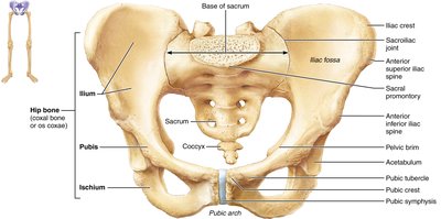
Bones of the Hip (Coxal Bones)
Each coxal bone is formed by the fusion of three bones: the ilium, ischium, and pubis. These bones meet at the acetabulum, a deep socket that receives the head of the femur, forming the hip joint.
Ilium: Superior region; articulates with the sacrum at the sacroiliac joint.
Ischium: Posteroinferior part; includes the ischial spine, lesser sciatic notch, and ischial tuberosity.
Pubis: Anterior portion; forms the pubic symphysis joint and the pubic arch.
Acetabulum: Deep socket formed at the fusion point; receives the femur head.


Pelvic Structure and Childbearing
Pelvic Brim, True and False Pelves
The pelvis is formed by the hip bones, sacrum, and coccyx. The pelvic brim (pelvic inlet) is a continuous oval ridge that separates the false pelvis (superior) from the true pelvis (inferior). The true pelvis defines the birth canal, and the pelvic outlet is the inferior margin of the true pelvis.
False pelvis: Superior to pelvic brim; supports abdominal organs.
True pelvis: Inferior to pelvic brim; contains pelvic organs and forms the birth canal.
Pelvic outlet: Bounded by pubic arch, ischia, sacrum, and coccyx.
Sexual Dimorphism of the Pelvis
There are distinct differences between male and female pelves, primarily related to childbearing. The female pelvis is wider, shallower, lighter, and rounder, adapted for childbirth. The male pelvis is narrower and deeper, adapted for supporting a heavier build and stronger muscles.
Characteristic | Female | Male |
|---|---|---|
General structure | Tilted forward; broad, shallow true pelvis | Tilted less; narrow, deep true pelvis |
Bone thickness | Lighter, thinner, smoother | Heavier, thicker, more prominent markings |
Acetabula | Smaller, farther apart | Larger, closer together |
Pubic arch/subpubic angle | Broader (80°–90°), more rounded | More acute (50°–60°) |
Sacrum | Wider, shorter, less curvature | Narrow, longer, more curvature |
Coccyx | More movable, projects inferiorly | Less movable, projects anteriorly |
Greater sciatic notch | Wide, shallow | Narrow, deep |
Pelvic inlet (brim) | Wider, oval | Narrow, heart-shaped |
Pelvic outlet | Wider, ischial spines farther apart | Narrower, ischial spines closer together |
The Lower Limb
Overview of Lower Limb Bones
The lower limb consists of three segments: the thigh, leg, and foot. It carries the entire weight of the erect body and is subjected to exceptional forces during activities such as jumping or running.
Thigh: Contains the femur, the largest and strongest bone in the body.
Leg: Composed of the tibia (medial, weight-bearing) and fibula (lateral, non-weight-bearing).
Foot: Includes tarsal bones, metatarsals, and phalanges.
Thigh – The Femur
The femur articulates proximally with the acetabulum of the hip and distally with the tibia and patella. Key features include:
Fovea capitis: Small pit in the head for ligament attachment.
Greater and lesser trochanters: Muscle attachment sites.
Intertrochanteric line and crest: Connect trochanters.
Gluteal tuberosity and linea aspera: Muscle attachment ridges.
Medial and lateral condyles: Articulate with tibia.
Patellar surface: Articulates with patella.
Intercondylar fossa: Between condyles.
Leg – Tibia and Fibula
The leg is made up of two parallel bones, the tibia and fibula, connected by an interosseous membrane. The tibia is the main weight-bearing bone, while the fibula is important for muscle attachment and stability.
Tibia: Medial, receives weight from femur, transmits to foot.
Fibula: Lateral, not weight-bearing, articulates with tibia.
Key features: Tibial condyles, intercondylar eminence, tibial tuberosity, medial malleolus, fibular notch; fibula head, lateral malleolus.
Foot – Tarsals, Metatarsals, and Phalanges
The foot skeleton includes the tarsus (7 tarsal bones), metatarsus (5 metatarsal bones), and phalanges (14 toe bones). The talus and calcaneus bear most of the body weight. The foot is structured to support weight and provide balance.
Tarsal bones: Talus, calcaneus, cuboid, navicular, medial/intermediate/lateral cuneiforms.
Metatarsals: Numbered I–V from hallux (big toe) to little toe; metatarsal I forms the ball of the foot.
Phalanges: Digit I (hallux) has two bones; digits II–V have three bones each.
Arches of the Foot
The foot has three arches maintained by interlocking bones, ligaments, and tendons. These arches allow the foot to bear weight and provide springiness for movement.
Lateral longitudinal arch: Low curve elevates lateral part of foot.
Medial longitudinal arch: Curves upward.
Transverse arch: Runs obliquely across the foot.
Developmental Aspects of the Skeleton
Infant Skull and Fontanelles
The infant skull has more bones than the adult skull, with unfused mandible and frontal bones. Skull bones are connected by fontanelles, which are unossified remnants of fibrous membranes. These allow for ease of birth and brain growth.
Fontanelles: Anterior, posterior, mastoid, sphenoidal.
Function: Allow skull deformation during birth and rapid brain growth.
Growth and Proportions
At birth, the cranium is large relative to the face. By 9 months, it reaches half adult size. Arms and legs grow faster than the head and trunk, leading to adult proportions.
Spinal Curvatures
Primary curvatures (thorax and sacrum) are convex at birth, resulting in a C-shaped spine. Secondary curvatures (cervical and lumbar) develop as the child grows, allowing upright posture.
Aging and Skeletal Changes
With age, intervertebral discs become thinner, less hydrated, and less elastic, increasing the risk of herniation. Height loss, ossification of costal cartilages, and bone mass reduction are common, increasing fracture risk.
Clinical Aspects
Bimalleolar Fracture
A bimalleolar fracture involves breaks in the distal ends of both the tibia and fibula, commonly seen in sports injuries.
Flat Foot (Pes Planus)
Flat foot may be congenital or acquired, caused by age, obesity, or high-impact activities. Proper footwear and orthotic inserts can help manage symptoms.
Cleft Palate
Cleft palate is a congenital abnormality where the right and left halves of the palate do not fuse, interfering with sucking and increasing the risk of aspiration pneumonia.
Developmental Dysplasia of the Hip (DDH)
DDH occurs in up to 2% of newborns, involving partial or complete dislocation of the femoral head and poorly developed acetabulum. High-risk infants include those with breech births. Early diagnosis and treatment are essential to prevent long-term complications.
