 뒤로
Chapter 8: Joints (Articulations) – Structure, Function, and Clinical Relevance
스터디 가이드 - 스마트 노트
 자료에 맞춘 맞춤형 노트, 핵심 정의, 예시, 맥락을 확장해 제공합니다.
Joints: An Overview
Definition and Importance
Joints, also known as articulations, are sites where two or more bones meet. They are essential for providing the skeleton with mobility and holding the skeleton together. The study of joints is called arthrology.
Structural classification: Based on the material binding bones together and the presence or absence of a joint cavity.
Functional classification: Based on the amount of movement allowed by the joint.

Classification of Joints
Structural Classification
Fibrous joints: Bones joined by dense fibrous connective tissue; no joint cavity.
Cartilaginous joints: Bones joined by cartilage; no joint cavity.
Synovial joints: Bones separated by a fluid-filled joint cavity.
Functional Classification
Synarthroses: Immovable joints
Amphiarthroses: Slightly movable joints
Diarthroses: Freely movable joints
Fibrous Joints
General Features
Fibrous joints are united by dense fibrous connective tissue and lack a joint cavity. Most are immovable (synarthroses), but some allow slight movement (amphiarthroses).
Types of Fibrous Joints
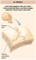
Sutures: Rigid, interlocking joints found only in the skull. Short connective tissue fibers allow for growth during youth; later ossify and fuse to protect the brain.
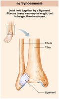
Syndesmoses: Bones connected by ligaments. Movement depends on fiber length—short fibers (e.g., inferior tibiofibular joint) allow little movement; longer fibers (e.g., interosseous membrane between radius and ulna) allow more movement.
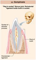
Gomphoses: Peg-in-socket joints, such as teeth in alveolar sockets. The periodontal ligament holds the tooth in place.
Cartilaginous Joints
General Features
Cartilaginous joints are united by cartilage and lack a joint cavity. They are either immovable or slightly movable.
Types of Cartilaginous Joints
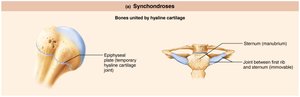
Synchondroses: Bones united by a bar or plate of hyaline cartilage. Most are immovable. Examples include the epiphyseal plates in children and the joint between the first rib and the manubrium of the sternum.
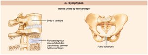
Symphyses: Bones united by fibrocartilage. These joints are strong and slightly movable. Examples include intervertebral joints and the pubic symphysis.
Synovial Joints
General Features
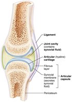
Synovial joints are characterized by the presence of a fluid-filled joint cavity. They are diarthroses (freely movable) and are the most common type of joint in the limbs.
Articular cartilage: Hyaline cartilage covering bone ends, preventing crushing.
Joint (articular) cavity: Small, fluid-filled space unique to synovial joints.
Articular capsule: Two layers—an external fibrous layer and an inner synovial membrane that produces synovial fluid.
Synovial fluid: Lubricates and nourishes articular cartilage; contains phagocytic cells.
Reinforcing ligaments: Strengthen and reinforce the joint.
Nerves and blood vessels: Detect pain, monitor joint position, and supply filtrate for synovial fluid.
Other Features
Bursae: Sacs of synovial fluid that reduce friction where ligaments, muscles, skin, tendons, or bones rub together.
Tendon sheaths: Elongated bursae that wrap around tendons subjected to friction (e.g., in the wrist).
Movements at Synovial Joints
Basic Principles
All muscles attach to bone or connective tissue at two or more points: the origin (immovable) and the insertion (movable). Muscle contraction moves the insertion toward the origin. Movements occur along transverse, frontal, or sagittal planes.
Range of Motion
Nonaxial: Slipping movements only
Uniaxial: Movement in one plane
Biaxial: Movement in two planes
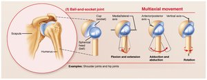
Multiaxial: Movement in or around all three planes
Types of Movements
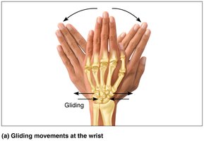
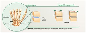
Gliding: One flat bone surface glides or slips over another. Example: intercarpal joints.
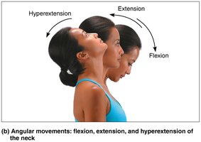
Angular movements: Change the angle between two bones.
Flexion: Decreases the angle of the joint
Extension: Increases the angle of the joint
Hyperextension: Extension beyond the anatomical position
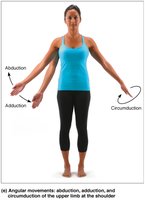
Abduction: Movement away from the midline
Adduction: Movement toward the midline
Circumduction: Distal end moves in a circle while the point of the cone is stationary
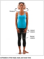
Rotation: Turning of a bone around its own long axis (e.g., rotation of the head, humerus, or femur).
Special Movements
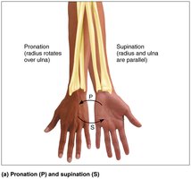
Supination and pronation: Rotation of the radius and ulna (forearm). Supination: palms face anteriorly; pronation: palms face posteriorly.
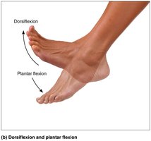
Dorsiflexion and plantar flexion: Upward and downward movement of the foot at the ankle.
Inversion and eversion: Turning the sole of the foot medially or laterally.
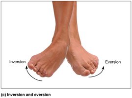
Protraction and retraction: Anterior and posterior movement in a transverse plane (e.g., mandible).
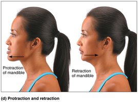
Elevation and depression: Lifting or lowering a body part (e.g., mandible).
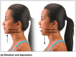
Opposition: Movement of the thumb to touch the tips of other fingers on the same hand.
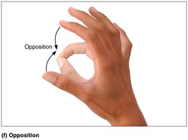
Types of Synovial Joints
Synovial joints are classified by the shape of their articular surfaces and the movements they allow.
Type | Movement | Example |
|---|---|---|
Plane | Nonaxial (gliding) | Intercarpal joints |
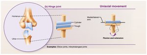
Hinge | Uniaxial (flexion/extension) | Elbow, interphalangeal joints |
Pivot | Uniaxial (rotation) | Proximal radioulnar joint, atlantoaxial joint |
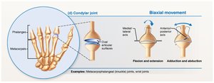
Condylar | Biaxial (flexion/extension, abduction/adduction) | Metacarpophalangeal joints |
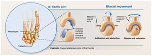
Saddle | Biaxial (flexion/extension, abduction/adduction) | Carpometacarpal joint of the thumb |
Ball-and-socket | Multiaxial (all movements) | Shoulder, hip |
The Knee Joint
Structure and Function
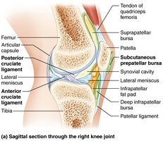
The knee is the largest and most complex joint in the body. It consists of three joints within a single cavity: the femoropatellar joint (plane joint) and the lateral and medial tibiofemoral joints (hinge joints).
Femoropatellar joint: Allows gliding motion during knee flexion.
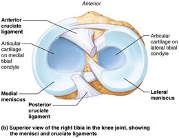
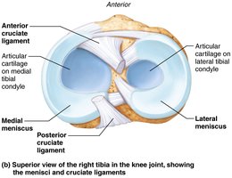
Tibiofemoral joints: Allow flexion, extension, and some rotation when the knee is partly flexed.
Ligaments and Stability
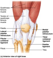
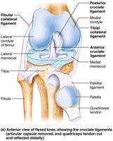
Extracapsular ligaments: Fibular and tibial collateral ligaments prevent rotation when the knee is extended.
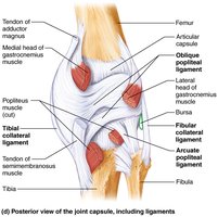
Intracapsular ligaments: Anterior cruciate ligament (ACL) prevents forward sliding of the tibia and stops hyperextension; posterior cruciate ligament prevents backward sliding of the tibia and forward sliding of the femur.
Bursae and Other Structures
About 12 bursae help reduce friction in the knee joint.
The quadriceps tendon gives rise to ligaments that stabilize the anterior aspect of the knee.
Common Joint Pathologies
Injuries
Sprains: Ligaments are stretched or torn.
Dislocations: Bones are forced out of alignment.
ACL tears: Common in athletes who change direction rapidly.
Joint cracking: Caused by pressure changes and air bubbles within the joint.
Arthritis
Osteoarthritis: Cartilage is broken down faster than it is replaced; bone spurs may form.
Rheumatoid arthritis: Autoimmune disease causing inflammation of the synovial membrane, destruction of tissue, and formation of scar tissue.
Check Your Understanding
Question: The articular joint capsule _____.
is a single layer of dense connective tissue
is characteristic of a gomphosis joint
is found in the interosseous membrane connecting the radius and ulna
is made up of hyaline cartilage
contains a membrane that makes synovial fluid (Correct answer)
