 뒤로
Chapter 8: Joints – Structured Study Notes for Anatomy & Physiology
스터디 가이드 - 스마트 노트
 자료에 맞춘 맞춤형 노트, 핵심 정의, 예시, 맥락을 확장해 제공합니다.
Joints: An Overview
Introduction to Joints
Joints, also known as articulations, are the sites where two or more bones meet. They are essential for providing mobility to the skeleton and holding it together. Understanding joints is crucial for diagnosing and treating injuries such as ankle sprains.
Classification of Joints
Structural Classification
Joints are classified structurally based on the material binding the bones and the presence or absence of a joint cavity. The three structural types are:
Fibrous Joints
Cartilaginous Joints
Synovial Joints
Functional Classification
Joints are also classified functionally based on the degree of movement they allow:
Synarthroses: Immovable joints
Amphiarthroses: Slightly movable joints
Diarthroses: Freely movable joints
Structural classification is more clear-cut and is commonly used in anatomical study.
Fibrous Joints
General Features
Fibrous joints are characterized by bones joined by dense fibrous connective tissue, with no joint cavity. Most are immovable, though mobility depends on the length of connective tissue fibers. There are three types:
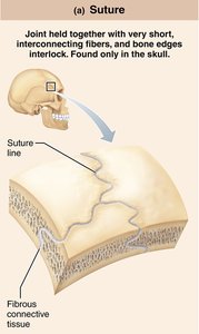
Sutures: Found only in the skull, held together by very short, interconnecting fibers; bone edges interlock.
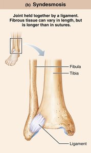
Syndesmoses: Bones connected by a ligament; fibrous tissue varies in length but is longer than in sutures.
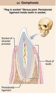
Gomphoses: "Peg-in-socket" joints, such as the periodontal ligament holding a tooth in its socket.
Cartilaginous Joints
General Features
Cartilaginous joints unite bones with cartilage and lack a joint cavity. They are not highly movable. Two main types exist:
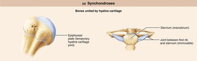
Synchondroses: Bones united by hyaline cartilage (e.g., epiphyseal plate, joint between first rib and sternum).
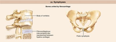
Symphyses: Bones united by fibrocartilage (e.g., intervertebral discs, pubic symphysis).
Synovial Joints
General Features
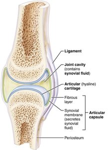
Synovial joints are characterized by a fluid-filled joint cavity separating the bones. All synovial joints are diarthrotic (freely movable) and include almost all limb joints. They possess six general features:
Articular cartilage: Hyaline cartilage covering bone ends, preventing crushing.
Joint (synovial) cavity: Unique, small, fluid-filled space.
Articular (joint) capsule: Two layers—external fibrous layer (dense irregular connective tissue) and inner synovial membrane (loose connective tissue producing synovial fluid).
Synovial fluid: Viscous, slippery filtrate of plasma and hyaluronic acid; lubricates and nourishes cartilage, contains phagocytic cells.
Reinforcing ligaments: Capsular, extracapsular, and intracapsular ligaments strengthen the joint.
Nerves and blood vessels: Nerves detect pain and monitor position; capillary beds supply filtrate for synovial fluid.
Bursae and Tendon Sheaths
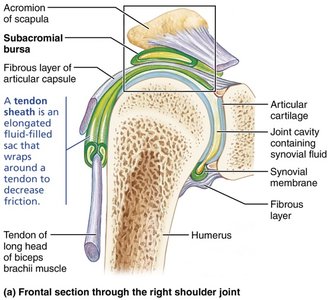
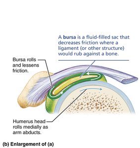
Bursae are fluid-filled sacs that decrease friction where ligaments or other structures rub against bone. Tendon sheaths are elongated bursae that wrap around tendons to reduce friction.
Factors Influencing Stability
Three factors determine the stability of synovial joints and help prevent dislocations:
Shape of articular surfaces: Shallow surfaces are less stable than ball-and-socket joints.
Ligament number and location: More ligaments provide greater strength.
Muscle tone: Most important factor; keeps tendons taut and reinforces joints, especially in the shoulder, knee, and foot arches.
Movements Allowed by Synovial Joints
Types of Movement
Synovial joints allow a range of motion, classified as:
Nonaxial: Gliding movements only
Uniaxial: Movement in one plane
Biaxial: Movement in two planes
Multiaxial: Movement in or around all three planes
Three general types of movements:
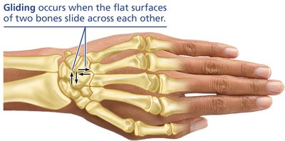
Gliding
Angular movements
Rotation
Gliding Movements
Gliding occurs when one flat bone surface slides over another. Examples include intercarpal and intertarsal joints, and between articular processes of vertebrae.
Angular Movements
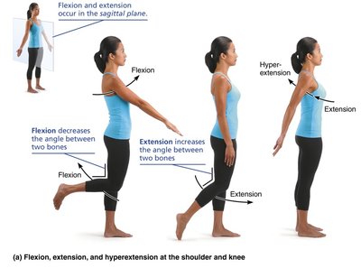
Angular movements change the angle between bones. Types include:
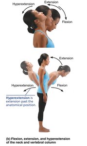
Flexion: Decreases the angle between bones, usually in the sagittal plane.
Extension: Increases the angle between bones, usually in the sagittal plane.
Hyperextension: Extension beyond anatomical position.
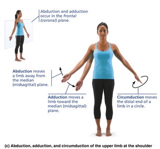
Abduction: Moves a limb away from the body midline in the frontal plane.
Adduction: Moves a limb toward the body midline in the frontal plane.
Circumduction: Moves a limb or finger so that it describes a cone in space.
Rotational Movements
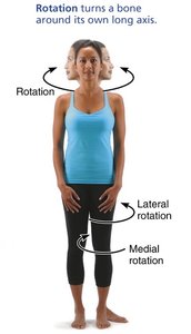
Rotation turns a bone around its own long axis. Types include:
Medial rotation: Rotating toward the median plane.
Lateral rotation: Rotating away from the median plane.
Special Movements
Some synovial joints allow special movements:
Pronation and supination: Rotational movements of the forearm.
Opposition: Movement of the thumb to touch other fingers.
Dorsiflexion and plantar flexion: Movements of the foot.
Inversion and eversion: Turning the sole of the foot medially or laterally.
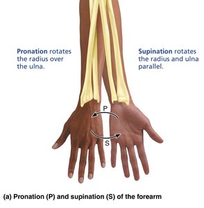
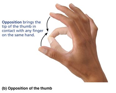
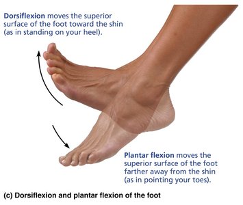
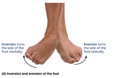
Table: Movements at Synovial Joints
Movement | Definition |
|---|---|
Gliding | Sliding the flat surfaces of two bones across each other |
Flexion | Decreasing the angle between two bones, usually in the sagittal plane |
Extension | Increasing the angle between two bones, usually in the sagittal plane |
Abduction | Moving a limb away from the body midline in the frontal plane |
Adduction | Moving a limb toward the body midline in the frontal plane |
Circumduction | Moving a limb or finger so that it describes a cone in space |
Rotation | Turning a bone around its longitudinal axis |
Medial rotation | Rotating toward the median plane |
Lateral rotation | Rotating away from the median plane |
Types of Synovial Joints
Classification by Shape and Movement
Synovial joints are classified into six types based on the shape of their articular surfaces and the movements they allow:
Plane
Hinge
Pivot
Condylar (Ellipsoid)
Saddle
Ball-and-socket
Selected Synovial Joints
Major Synovial Joints
Some synovial joints have unique structural features, abilities, and weaknesses. The five main synovial joints are:
Temporomandibular (Jaw) Joint
Shoulder Joint
Elbow Joint
Hip Joint
Knee Joint
Clinical Relevance
Dislocations and injuries are common in certain joints due to their structural features:
Temporomandibular Joint (TMJ): Shallow socket makes dislocation common, usually anteriorly. Symptoms include pain, stiffness, and popping sounds. Treatment may involve realignment and bite plates.
Shoulder Joint: High mobility makes dislocations common, especially anteriorly and inferiorly. Reinforcing tendons (rotator cuff) are crucial for stability.
Knee Joint: Vulnerable to horizontal blows; common injuries involve collateral ligaments, cruciate ligaments, and menisci. The "unhappy triad" refers to simultaneous injury of the ACL, tibial collateral ligament, and meniscus.
Example: A football player may dislocate the shoulder when tackling, due to poor support from the glenoid cavity.
Additional info: Surgery is often required for severe ligament or meniscus injuries in the knee.
Summary Table: Structural and Functional Characteristics of Body Joints
Joint Type | Binding Material | Joint Cavity | Movement |
|---|---|---|---|
Fibrous | Dense fibrous connective tissue | No | Mostly immovable |
Cartilaginous | Cartilage | No | Slightly movable |
Synovial | Articular cartilage, synovial fluid | Yes | Freely movable |
