 뒤로
뒤로Chapter 8: Special Senses – Anatomy & Physiology Study Notes
스터디 가이드 - 스마트 노트
 자료에 맞춘 맞춤형 노트, 핵심 정의, 예시, 맥락을 확장해 제공합니다.
Special Senses Overview
Definition and Classification
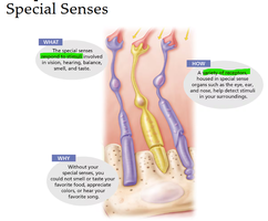
The special senses are specialized sensory systems responsible for vision, hearing, balance (equilibrium), smell, and taste. These senses are distinct from the general senses (touch, temperature, pain, proprioception) and rely on complex organs or localized clusters of receptors.
General senses: Touch, temperature, pain, proprioception (muscles and joints)
Special senses: Smell, taste, sight, hearing, equilibrium
Special sense receptors: Large, complex organs (eye, ear) or clusters (taste buds, olfactory epithelium)
Example: The nervous system integrates sensory input from these receptors to produce motor output and interpret the environment.
The Eye and Vision
Accessory Structures of the Eye
The eye is protected and supported by several accessory structures, which include muscles, glands, and membranes.
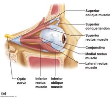
Extrinsic eye muscles: Six muscles attach to the outer surface, producing gross eye movements.
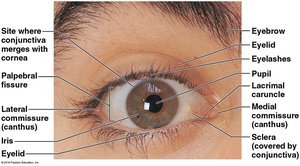
Eyelids: Meet at the medial and lateral commissures (canthus); protect the eye.
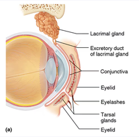
Eyelashes: Tarsal glands produce oily secretions; ciliary glands are located between lashes.
Conjunctiva: Membrane lining eyelids and eyeball; connects with cornea; secretes mucus for lubrication.
Lacrimal apparatus: Lacrimal gland produces tears; ducts drain tears into nasal cavity.
Example: Tears contain salt, mucus, antibodies, and lysozyme, serving to cleanse, protect, moisten, and lubricate the eye.
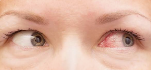
Homeostatic Imbalance: Conjunctivitis
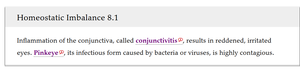
Conjunctivitis is inflammation of the conjunctiva, resulting in red, irritated eyes. The infectious form, known as pinkeye, is highly contagious and caused by bacteria or viruses.
Extrinsic Eye Muscles
Six extrinsic muscles control the movement of the eyeball, allowing for coordinated and precise visual tracking.
Superior, inferior, lateral, and medial rectus muscles
Superior and inferior oblique muscles
Internal Structures of the Eyeball
The eyeball is composed of three main layers (tunics) and is filled with fluids called humors. The lens divides the eye into anterior and posterior chambers.
Fibrous layer: Sclera (white of the eye) and cornea (transparent, allows light entry)
Vascular layer: Choroid (nutritive, pigmented), ciliary body (smooth muscle), iris (regulates light, gives eye color), pupil (opening in iris)
Sensory layer: Retina (outer pigmented layer absorbs light; inner neural layer contains photoreceptors: rods and cones)
Example: The cornea is the only human tissue that can be transplanted without fear of rejection due to its lack of blood vessels.
Retina and Photoreceptors
The retina contains two types of photoreceptors:
Rods: Peripheral vision, dim light, gray tones
Cones: Color vision, sharpest vision at fovea centralis
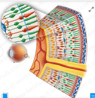
Example: The optic disc (blind spot) is where the optic nerve exits the eye; no photoreceptors are present here.
Lens and Humors
The lens is a flexible, biconvex structure that focuses light onto the retina. The eye contains two types of humors:
Aqueous humor: Watery fluid in anterior chamber; maintains intraocular pressure, provides nutrients
Vitreous humor: Gel-like substance in posterior chamber; prevents eye collapse, maintains pressure
Physiology of Vision
Light is refracted by the cornea, lens, and humors to focus on the retina. The image formed is real, reversed left to right, upside down, and smaller than the object.
Accommodation: Lens changes shape to focus on near objects
Visual pathway: Optic nerve → optic chiasma → optic tract → thalamus → optic radiation → visual cortex (occipital lobe)
Example: Binocular vision provides depth perception (three-dimensional vision).
Common Vision Disorders
Emmetropia: Normal focusing
Myopia (nearsightedness): Distant objects blurry; eyeball too long
Hyperopia (farsightedness): Near objects blurry; eyeball too short or lazy lens
Astigmatism: Blurry images due to unequal curvatures of cornea or lens
The Ear: Hearing and Balance
Anatomy of the Ear
The ear is divided into three regions, each with distinct functions:
External (outer) ear: Auricle (pinna), external acoustic meatus, tympanic membrane; collects sound waves
Middle ear: Air-filled cavity; contains ossicles (malleus, incus, stapes); transmits and amplifies vibrations
Internal (inner) ear: Bony labyrinth (cochlea, vestibule, semicircular canals); contains perilymph and endolymph; houses receptors for hearing and equilibrium
Hearing
Sound waves are transmitted through the ear, causing vibrations that stimulate hair cells in the cochlea. The cochlear nerve carries impulses to the auditory cortex.
High-pitched sounds: Stimulate short, stiff fibers near oval window
Low-pitched sounds: Stimulate long, floppy fibers further along cochlea
Equilibrium
Equilibrium is maintained by the vestibular apparatus in the inner ear, which has two functional parts:
Static equilibrium: Maculae in vestibule report head position; otoliths bend hair cells
Dynamic equilibrium: Crista ampullaris in semicircular canals responds to rotational movements; cupula bends hair cells
Hearing and Equilibrium Deficits
Conduction deafness: Sound transmission hindered in external/middle ear
Sensorineural deafness: Damage to nervous system structures
Ménière’s syndrome: Inner ear disorder causing progressive deafness and vertigo
Chemical Senses: Smell and Taste
Olfactory Receptors and Smell
Olfactory receptors are located in the roof of the nasal cavity. Chemicals must be dissolved in mucus to be detected by olfactory hairs (cilia).
Taste Buds and the Sense of Taste
Taste buds are found on the tongue, soft palate, pharynx, and cheeks. The tongue is covered with papillae containing taste buds.
Vallate (circumvallate) papillae
Fungiform papillae
Filiform papillae
Gustatory cells possess gustatory hairs that are stimulated by chemicals dissolved in saliva.
Sweet: Sugars, saccharine, some amino acids
Sour: Ions or acids
Bitter: Alkaloids
Salty: Metal ions
Umami: Glutamate, beefy taste
Developmental Aspects of the Special Senses
Embryonic Development and Age-Related Changes
Special sense organs form early in embryonic development. Maternal infections can cause visual and auditory abnormalities. Vision requires the most learning; infants are farsighted and lack color vision and depth perception. The eye matures until age 8 or 9.
Presbyopia: Age-related decrease in lens elasticity; difficulty focusing on close objects
Presbycusis: Age-related sensorineural deafness; may result from otosclerosis (ossicle fusion)
Taste and smell: Most acute at birth; sensitivity decreases after age 40
Example: Congenital ear problems may result from missing pinnas or external acoustic meatuses.
Special Sense | Main Receptor Location | Type of Receptor |
|---|---|---|
Vision | Retina (eye) | Photoreceptors (rods, cones) |
Hearing | Cochlea (ear) | Mechanoreceptors (hair cells) |
Equilibrium | Vestibular apparatus (ear) | Mechanoreceptors (maculae, crista ampullaris) |
Smell | Olfactory epithelium (nose) | Chemoreceptors |
Taste | Taste buds (tongue, palate, pharynx) | Chemoreceptors |
Additional info: The notes above expand on brief points from the original materials, providing definitions, examples, and academic context for each special sense.
