 뒤로
Chapter 8A: Joints – Structure, Function, and Classification
스터디 가이드 - 스마트 노트
 자료에 맞춘 맞춤형 노트, 핵심 정의, 예시, 맥락을 확장해 제공합니다.
Joints: Structure, Function, and Classification
Introduction to Joints
Joints, also known as articulations, are the sites where two or more bones meet. They are essential for providing mobility to the skeleton and holding it together. Understanding joint structure and function is crucial for diagnosing and treating musculoskeletal injuries.
Classification of Joints
Joints are classified in two main ways: structurally and functionally.
Structural Classification: Based on the material binding the bones and the presence or absence of a joint cavity. The three types are:
Fibrous Joints
Cartilaginous Joints
Synovial Joints
Functional Classification: Based on the degree of movement allowed:
Synarthroses: Immovable joints
Amphiarthroses: Slightly movable joints
Diarthroses: Freely movable joints
Fibrous Joints
General Structure of Fibrous Joints
Fibrous joints are characterized by bones joined by dense fibrous connective tissue. They lack a joint cavity and are mostly immovable, though the degree of movement depends on the length of the connective tissue fibers.
Three main types: Sutures, Syndesmoses, and Gomphoses
Sutures
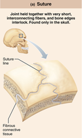
Sutures are rigid, interlocking joints found only in the skull. They allow for growth during youth and contain short connective tissue fibers that permit expansion. In middle age, sutures ossify and fuse, forming immovable joints called synostoses that protect the brain.
Example: Sutures between skull bones
Syndesmoses
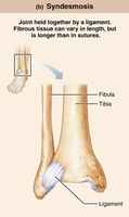
Syndesmoses are joints where bones are connected by ligaments, which are bands of fibrous tissue. The length of the fibers determines the degree of movement. Short fibers offer little to no movement (e.g., inferior tibiofibular joint), while longer fibers allow more movement (e.g., interosseous membrane between radius and ulna).
Example: Inferior tibiofibular joint (little movement), interosseous membrane (more movement)
Gomphoses
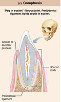
Gomphoses are peg-in-socket fibrous joints. The only examples are the teeth in their alveolar sockets, where the fibrous connection is the periodontal ligament that holds the tooth in place.
Example: Tooth in alveolar socket
Cartilaginous Joints
General Structure of Cartilaginous Joints
Cartilaginous joints are formed by bones united by cartilage. Like fibrous joints, they lack a joint cavity and are not highly movable. There are two main types: Synchondroses and Symphyses.
Synchondroses
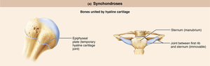
Synchondroses unite bones with a bar or plate of hyaline cartilage. Most are synarthrotic (immovable). Examples include temporary epiphyseal plate joints (which become synostoses after closure) and the joint between the first rib and the manubrium of the sternum.
Example: Epiphyseal plate, first rib-sternum joint
Symphyses
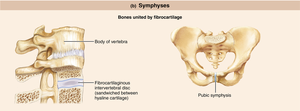
Symphyses unite bones with fibrocartilage, with hyaline cartilage present as articular cartilage on bony surfaces. These joints are strong and amphiarthrotic (slightly movable). Examples include intervertebral joints and the pubic symphysis.
Example: Intervertebral discs, pubic symphysis
Synovial Joints
General Structure of Synovial Joints
Synovial joints are characterized by a fluid-filled joint cavity that separates the bones. All synovial joints are diarthrotic (freely movable) and include almost all limb joints. They have six general features, bursae and tendon sheaths, and their stability is influenced by three main factors.
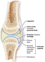
Articular cartilage: Hyaline cartilage covers bone ends, preventing crushing.
Joint (synovial) cavity: Small, fluid-filled space unique to synovial joints.
Articular (joint) capsule: Two layers thick, enclosing the joint.
Synovial fluid: Viscous, slippery filtrate of plasma and hyaluronic acid; lubricates and nourishes cartilage, contains phagocytic cells.
Reinforcing ligaments: Capsular (thickened part of fibrous layer), extracapsular (outside capsule), intracapsular (deep to capsule).
Nerves and blood vessels: Nerves detect pain and monitor position/stretch; capillary beds supply filtrate for synovial fluid.
Other Features of Synovial Joints
Fatty pads: Cushioning between fibrous layer and synovial membrane or bone.
Articular discs (menisci): Fibrocartilage separates articular surfaces to improve fit, stabilize joint, and reduce wear and tear.
Bursae and Tendon Sheaths
Bursae are bags of synovial fluid that act as lubricating ball bearings, reducing friction where ligaments, muscles, skin, tendons, or bones rub together. Tendon sheaths are elongated bursae wrapped around tendons subjected to friction.
Bursae: Not strictly part of synovial joints but closely associated.
Tendon sheaths: Elongated bursae around tendons.
Factors Influencing Stability of Synovial Joints
Three factors determine the stability of synovial joints and help prevent dislocations:
Shape of articular surfaces: Shallow surfaces are less stable than ball-and-socket joints (minor role).
Ligament number and location: More ligaments provide greater strength (limited role).
Muscle tone: Most important factor; keeps tendons taut as they cross joints, especially important in shoulder, knee, and foot arches.
Summary Table: Structural and Functional Classification of Joints
Structural Type | Material | Joint Cavity? | Functional Type | Examples |
|---|---|---|---|---|
Fibrous | Dense fibrous connective tissue | No | Synarthroses (mostly immovable) | Sutures, syndesmoses, gomphoses |
Cartilaginous | Cartilage (hyaline or fibrocartilage) | No | Synarthroses or amphiarthroses | Synchondroses, symphyses |
Synovial | Articular cartilage, synovial fluid | Yes | Diarthroses (freely movable) | Most limb joints |
Example: The knee joint is a synovial joint, stabilized by ligaments and muscle tone, and contains bursae to reduce friction.
Additional info: Academic context was added to clarify the structural and functional classification, as well as the importance of joint stability factors.
