 뒤로
Chapter 9: The Muscular System – Structure, Function, and Physiology
스터디 가이드 - 스마트 노트
 자료에 맞춘 맞춤형 노트, 핵심 정의, 예시, 맥락을 확장해 제공합니다.
The Muscular System
Functions of the Muscular System
The muscular system is essential for movement, posture, joint stabilization, temperature regulation, and controlling the movement of substances between body compartments.
Movement: Muscles contract to produce movement of the body and its parts.
Maintaining posture: Continuous muscle contractions keep the body upright.
Stabilizing joints: Muscles reinforce and stabilize joints during movement.
Temperature homeostasis: Muscle activity generates heat, helping maintain body temperature.
Regulation of substances: Muscles control passage of substances, such as food through the digestive tract.
Characteristics of Muscle Tissue
Muscle tissue possesses unique properties that enable its function:
Excitability: Ability to receive and respond to stimuli.
Contractility: Ability to shorten forcibly when stimulated.
Extensibility: Ability to be stretched beyond resting length.
Elasticity: Ability to return to original length after stretching.
Muscle Types
Skeletal, Cardiac, and Smooth Muscle
There are three main types of muscle tissue, each with distinct structure and function:
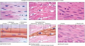
Skeletal Muscle: Voluntary, striated, attached to bones, multinucleated, limited regeneration.
Cardiac Muscle: Involuntary, striated, found in heart walls, single nucleus, no regeneration.
Smooth Muscle: Involuntary, non-striated, found in walls of hollow organs, single nucleus, fast regeneration.
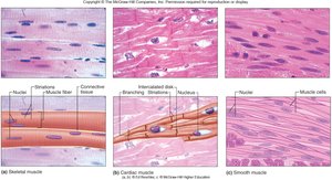
Skeletal Muscle Structure
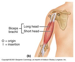
Muscle Attachments: Origin and Insertion
Skeletal muscles attach to bones at two points:
Origin: Attachment to the bone that remains stationary during contraction.
Insertion: Attachment to the bone that moves during contraction.
Direct vs. Indirect Attachment: Muscles may attach directly via the epimysium or indirectly via tendons or aponeuroses.
Levels of Organization
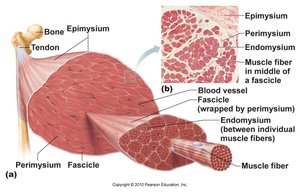
Skeletal muscle is organized into hierarchical structures:
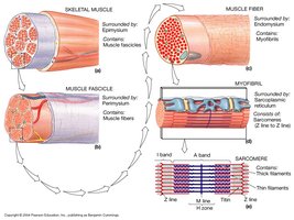
Muscle: Surrounded by epimysium.
Fascicle: Bundle of muscle fibers, surrounded by perimysium.
Muscle Fiber: Individual muscle cell, surrounded by endomysium.
Myofibril: Rod-like structure within muscle fiber, contains contractile proteins.
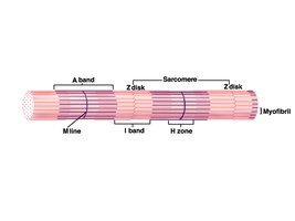
Sarcomere: Functional unit of contraction, composed of thick (myosin) and thin (actin) filaments.
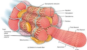
Microscopic Anatomy: Sarcomere
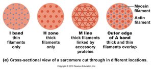
The sarcomere is the basic contractile unit of skeletal muscle, defined by the arrangement of thick and thin filaments:
A band: Contains both myosin and actin.
H zone: Contains only myosin (thick filaments).
M line: Middle of the sarcomere, bisects H zone.
I band: Contains only actin (thin filaments).
Z disc: Boundary of the sarcomere, anchors thin filaments.
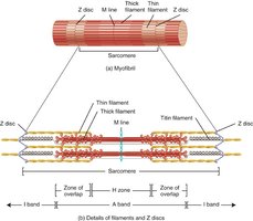
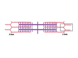
Thick and Thin Filaments
Thick filaments are composed of myosin, while thin filaments are primarily actin:
Myosin: Double-headed protein, forms cross-bridges, contains actin and ATP binding sites.
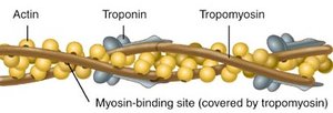
Actin: Two strands wound together, contains myosin binding sites.
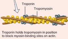
Tropomyosin: Covers myosin binding sites on actin when muscle is at rest.
Troponin: Binds calcium, moves tropomyosin to expose binding sites for contraction.
Muscle Contraction: Sliding Filament Mechanism
Mechanism of Contraction
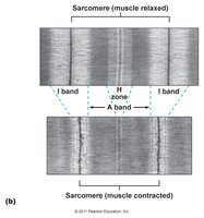
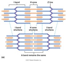
Muscle contraction occurs when thick and thin filaments slide past each other, shortening the sarcomere:
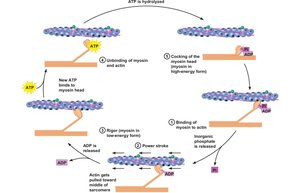
Activation of cross-bridges: Myosin heads bind to actin, forming cross-bridges.
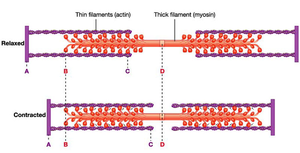
Sliding: Filaments slide, but do not shorten themselves.
Band changes: A band remains constant, I band and H zone shorten.
Excitation-Contraction Coupling
This process links the electrical signal from a motor neuron to muscle contraction:
Action potential: Travels along sarcolemma and T-tubules.
Calcium release: Sarcoplasmic reticulum releases Ca2+ into cytoplasm.
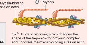
Troponin activation: Ca2+ binds to troponin, moving tropomyosin and exposing myosin binding sites.
Cross-bridge formation: Myosin heads bind to actin, initiating contraction.
Muscle Physiology
Ion Channels and Pumps
Muscle cells use ion channels and pumps to maintain membrane potential and respond to stimulation:
Ion Channels: Allow ions to diffuse across the membrane.
Gated Channels: Open in response to voltage or ligand binding.
Pumps: Move ions against concentration gradients, require ATP.
Resting membrane potential: Typically -70 mV in muscle cells.
Nerve Stimulation and Neuromuscular Junction
Each muscle cell is stimulated by a motor neuron at the neuromuscular junction:
Structure: Axon end bulb contains acetylcholine (ACh), synaptic cleft, motor end plate with ACh receptors.
Process: Nerve impulse releases ACh, which binds to receptors, causing Na+ influx and depolarization.
Motor Units and Muscle Response
Motor Units
A motor unit consists of a motor neuron and all the muscle fibers it innervates:
Size: Can range from a few to several hundred muscle fibers per neuron.
Function: All fibers in a motor unit contract together; smaller units allow finer control.
All-or-None Principle and Graded Response
Individual muscle fibers contract fully when stimulated, but whole muscles can vary force and duration:
All-or-None: Each fiber contracts to its full extent.
Graded Response: Muscle force depends on number and frequency of motor units activated.
Energy for Muscle Contraction
ATP and Energy Sources
ATP is the direct energy source for muscle contraction, but is stored only briefly:
Creatine Phosphate: Regenerates ATP for short bursts (15 seconds).
Anaerobic Respiration: Produces ATP quickly, yields lactic acid, supports activity up to 45 seconds.
Aerobic Respiration: Requires oxygen, produces large amounts of ATP, supports prolonged activity.
Muscle Fatigue and Oxygen Debt
Muscle fatigue is the inability to contract despite stimulation, often due to metabolic imbalances. Oxygen debt is the amount of oxygen needed to restore muscle cells after activity.
Muscle Cell Types
Slow Oxidative, Fast Glycolytic, and Fast Oxidative Fibers
Muscles contain a mix of fiber types, each suited for different activities:
Slow Oxidative (Type I): Thin, endurance, aerobic, abundant myoglobin and mitochondria.
Fast Glycolytic (Type IIb): Large diameter, powerful, anaerobic, fatigues quickly, little myoglobin.
Fast Oxidative (Type IIa): Intermediate, aerobic and anaerobic, fatigue resistant, intermediate power.
Effect of Exercise and Disorders
Exercise Effects
Regular exercise increases muscle fiber size and ATP production capacity. Disuse or nerve damage leads to atrophy.
Muscle Disorders
Muscle Cramps: Involuntary contractions, often due to electrolyte imbalance.
Muscular Dystrophy: Genetic disease causing muscle degeneration, most commonly Duchenne muscular dystrophy.
Polio: Viral disease destroying motor neurons.
Smooth Muscle
Gross and Microscopic Anatomy
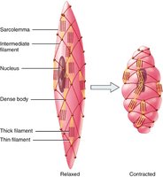
Smooth muscle is found in walls of hollow organs, arranged in layers, and contracts via peristalsis. Cells are spindle-shaped, have one nucleus, and lack sarcomeres.
Contraction Mechanism
Smooth muscle contracts via actin and myosin, but uses a different mechanism than skeletal muscle. Contractions are slow, synchronized, and resistant to fatigue.
Muscle Type | Control | Striations | Nuclei | Regeneration |
|---|---|---|---|---|
Skeletal | Voluntary | Striated | Many | Limited |
Cardiac | Involuntary | Striated | One | None |
Smooth | Involuntary | Non-striated | One | Fast |
Additional info: Fiber types, contraction mechanisms, and energy metabolism are influenced by genetics and training. Disorders such as muscular dystrophy and polio highlight the importance of muscle structure and function in health.
