 뒤로
Classification and Function of Joints in Human Anatomy
스터디 가이드 - 스마트 노트
 자료에 맞춘 맞춤형 노트, 핵심 정의, 예시, 맥락을 확장해 제공합니다.
Joints: Classification and Function
Overview of Joint Classification
Joints, also known as articulations, are points of contact between bones that allow for movement and provide structural stability. They are classified both structurally and functionally, which determines their mobility and anatomical features.
Structural Classification: Based on the material binding bones and presence/absence of a joint cavity.
Functional Classification: Based on the degree of movement permitted.
Structural and Functional Types of Joints
Type | Structural Features | Functional Classification | Examples |
|---|---|---|---|
Fibrous Joints | Bones fit closely together, joined by fibrous tissue | Synarthroses (Immovable) | Sutures, syndesmoses, gomphoses |
Cartilaginous Joints | Bones joined by hyaline cartilage or fibrocartilage | Amphiarthroses (Slightly movable) | Synchondroses, symphyses |
Synovial Joints | Fluid-filled joint capsule between bones | Diarthroses (Freely movable) | Most joints in the body |
Fibrous Joints (Synarthroses)
Characteristics and Types
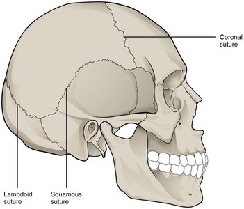
Fibrous joints are immovable and the bones are tightly joined by fibrous connective tissue. They are primarily found in areas requiring stability.
Sutures: Found only in the skull; interlocking projections provide strength.
Syndesmoses: Ligaments connect two bones, e.g., distal tibiofibular joint.
Gomphoses: Peg-in-socket joints, such as the connection between a tooth and its socket.
Cartilaginous Joints (Amphiarthroses)
Characteristics and Types
Cartilaginous joints allow limited movement and are joined by cartilage. They are important for flexibility and shock absorption.
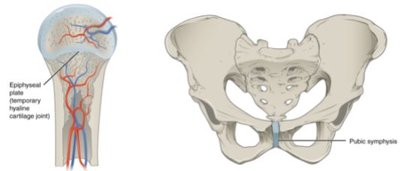
Symphyses: Pad or disk of fibrocartilage between bones, e.g., intervertebral discs, pubic symphysis.
Synchondroses: Hyaline cartilage between bones, e.g., epiphyseal plates, joints between ribs and sternum.
Synovial Joints (Diarthroses)
Structure and Components
Synovial joints are freely movable and have a complex structure that allows for a wide range of motion. They are the most common type of joint in the body.
Ligaments: Dense connective tissue that stabilizes the joint.
Synovial membrane: Lines the joint capsule and secretes synovial fluid.
Articular capsule: Encloses the joint cavity.
Articular cartilage: Hyaline cartilage covering bone surfaces.
Bursae: Fluid-filled sacs that cushion and reduce friction.
Synovial cavity: Space containing synovial fluid for lubrication.


Types of Synovial Joints
Classification by Movement
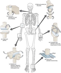
Synovial joints are classified by the number of axes around which movement occurs:
Uniaxial: Movement in one plane (e.g., hinge, pivot joints).
Biaxial: Movement in two planes (e.g., saddle, condyloid joints).
Multiaxial: Movement in three or more planes (e.g., ball-and-socket, plane joints).
Joint Stability and Movement
Relationship Between Stability and Mobility
Joints balance stability and mobility. Highly stable joints allow little movement, while highly mobile joints are less stable. Range of motion (ROM) is used to assess joint function and injury.
Stable joints: Limited mobility, e.g., sutures in the skull.
Mobile joints: Greater movement, e.g., shoulder joint.
ROM assessment: Measured actively or passively, often using a goniometer.

Movements at Joints
Basic Movements
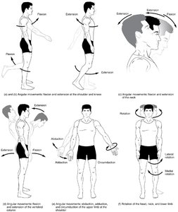
Joints allow for a variety of movements essential for daily activities and complex tasks. These movements are classified as angular, rotational, or special movements.
Extension: Increases the angle between bones, straightening the joint.
Flexion: Decreases the angle, bending the joint.
Rotation: Pivoting a bone on its axis.
Circumduction: Circular movement of a limb's distal end.
Abduction: Moves a part away from the body's midline.
Adduction: Moves a part toward the body's midline.

Special Movements
Some joints allow unique movements, particularly in the hands, feet, and jaw.
Supination: Turns the palm upward.
Pronation: Turns the palm downward.
Plantar flexion: Increases angle between top of foot and leg.
Dorsiflexion: Decreases angle between top of foot and leg.
Inversion: Turns sole of foot inward.
Eversion: Turns sole of foot outward.
Protraction: Moves a part forward.
Retraction: Moves a part backward.
Elevation: Moves a part up.
Depression: Lowers a part.
Opposition: Brings thumb tip in contact with a finger tip.
Interactive Activities and Further Study
Resources for Learning
To reinforce understanding, students are encouraged to use interactive activities, visual books, and textbook readings. These resources provide practice questions, animations, and detailed explanations of joint anatomy and physiology.
Interactive activities: Joints interactives, MCQs, anatomy drills.
Visual books: Joints and ligaments coloring books.
Readings: Textbook chapters and summaries from OpenStax, Boundless, CliffsNotes.
Short clips: Animations and video lectures on joint types and movements.
Summary Table: Joint Types and Examples
Joint Type | Movement | Example |
|---|---|---|
Fibrous (Synarthroses) | Immovable | Sutures in skull |
Cartilaginous (Amphiarthroses) | Slightly movable | Intervertebral discs, pubic symphysis |
Synovial (Diarthroses) | Freely movable | Shoulder, knee, hip joints |
Key Terms: Joint, articulation, synarthroses, amphiarthroses, diarthroses, suture, syndesmosis, gomphosis, symphysis, synchondrosis, synovial membrane, articular capsule, bursae, range of motion (ROM), goniometer.
