 뒤로
뒤로Comprehensive Anatomy & Physiology I Study Guidance
스터디 가이드 - 스마트 노트
 자료에 맞춘 맞춤형 노트, 핵심 정의, 예시, 맥락을 확장해 제공합니다.
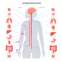
Q1. Define/Describe: Visceral Reflexes, Structure and Function of the Autonomic Nervous System (ANS), Structure & Function of Sympathetic and Parasympathetic Divisions.
Background
Topic: Autonomic Nervous System (ANS)
This question tests your understanding of the ANS, including its divisions (sympathetic and parasympathetic), their structure, function, and the concept of visceral reflexes.
Key Terms:
Visceral Reflexes: Involuntary responses of internal organs to stimuli, mediated by the ANS.
ANS: A division of the peripheral nervous system that controls involuntary functions (e.g., heart rate, digestion).
Sympathetic Division: Prepares the body for 'fight or flight' responses.
Parasympathetic Division: Promotes 'rest and digest' activities.
Key Concepts:
Sympathetic: Short preganglionic, long postganglionic fibers; uses norepinephrine; increases heart rate, dilates pupils.
Parasympathetic: Long preganglionic, short postganglionic fibers; uses acetylcholine; decreases heart rate, stimulates digestion.
Step-by-Step Guidance
Start by defining visceral reflexes and their role in maintaining homeostasis.
Describe the overall structure of the ANS, including its two main divisions.
Compare the anatomical differences between sympathetic and parasympathetic pathways (fiber length, neurotransmitters).
Explain the functional effects of each division on target organs (e.g., heart, lungs, digestive tract).
Review the diagram to identify which organs are innervated by each division and how their activity changes.
Try solving on your own before revealing the answer!
Final Answer:
Visceral reflexes are automatic, involuntary responses of internal organs, regulated by the ANS. The ANS consists of the sympathetic (fight or flight) and parasympathetic (rest and digest) divisions. The sympathetic division has short preganglionic and long postganglionic fibers, uses norepinephrine, and increases heart rate and energy expenditure. The parasympathetic division has long preganglionic and short postganglionic fibers, uses acetylcholine, and promotes maintenance functions like digestion and decreased heart rate. The diagram shows how each division innervates various organs and modulates their activity.
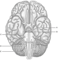
Q2. Using Figure 13.1, match the following cranial nerves to their functions.
Background
Topic: Cranial Nerves
This question tests your ability to identify cranial nerves and their functions using anatomical diagrams.
Key Terms:
Cranial Nerves: Twelve pairs of nerves that emerge directly from the brain.
Superior Oblique Muscle: Eye muscle innervated by the trochlear nerve.
Vagus Nerve: Longest cranial nerve, involved in parasympathetic control.
Step-by-Step Guidance
Review the diagram and locate the labeled cranial nerves.
Recall the primary functions of each cranial nerve (e.g., movement, sensation, autonomic control).
Match each nerve to its described function (e.g., which nerve innervates the superior oblique muscle).
Consider clinical consequences of nerve damage (e.g., loss of balance, speech difficulties).
Try solving on your own before revealing the answer!
Final Answer:
1) Trochlear nerve (innervates superior oblique muscle), 2) Vagus nerve (longest cranial nerve), 3) Vestibulocochlear nerve (damage causes dizziness, nausea, loss of balance), 4) Vagus nerve (involved in digestive tract movement), 5) Hypoglossal nerve (damage causes speech/swallowing difficulty), 6) Trochlear nerve (damage prevents inferolateral eye rotation).
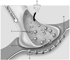
Q3. Using Figure 11.2, match the following structures at the neuromuscular junction.
Background
Topic: Neuromuscular Junction
This question tests your understanding of the anatomy and physiology of the neuromuscular junction.
Key Terms:
Synaptic Vesicles: Store neurotransmitters in the axon terminal.
Ion Channels: Allow ions to pass through membranes.
Synaptic Cleft: Space between neuron and muscle cell.
Postsynaptic Membrane: Receives neurotransmitter signal.
Calcium Ions: Trigger neurotransmitter release.
Step-by-Step Guidance
Identify each labeled structure in the diagram (A-E).
Recall the function of each structure (e.g., what triggers neurotransmitter release).
Match the description to the correct label (e.g., ion channel, synaptic vesicle).
Review the sequence of events during synaptic transmission.
Try solving on your own before revealing the answer!
Final Answer:
9) E: Ion channel, 10) C: Synaptic vesicles, 11) A: Calcium ions, 12) B: Postsynaptic membrane, 13) D: Synaptic cleft.
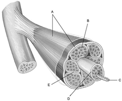
Q4. Using Figure 9.1, match the following muscle structures.
Background
Topic: Skeletal Muscle Anatomy
This question tests your ability to identify and describe the connective tissue components of skeletal muscle.
Key Terms:
Endomysium: Surrounds individual muscle fibers.
Perimysium: Surrounds bundles of muscle fibers (fascicles).
Epimysium: Covers the entire muscle organ.
Muscle Fiber: Single muscle cell.
Step-by-Step Guidance
Examine the diagram and locate each labeled structure.
Recall the function and location of each connective tissue layer.
Match the description to the correct label (e.g., which sheath surrounds individual fibers).
Review the hierarchical organization of muscle tissue.
Try solving on your own before revealing the answer!
Final Answer:
1) E: Endomysium, 2) B: Fascicle, 3) A: Epimysium, 4) D: Perimysium, 5) C: Muscle fiber.
Q5. Using Figure 7.1, match the following vertebral column regions and functions.
Background
Topic: Vertebral Column Anatomy
This question tests your ability to identify regions of the vertebral column and their anatomical features.
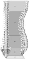
Key Terms:
Cervical Curvature: Uppermost region, includes atlas and axis.
Thoracic Curvature: Attaches to ribs.
Lumbar Curvature: Bears most of the weight.
Sacral and Coccyx: Fused vertebrae at the base.
Step-by-Step Guidance
Review the diagram and identify each labeled region (A-E).
Recall the anatomical features and functions of each region.
Match the description to the correct label (e.g., which region bears most weight).
Consider clinical relevance (e.g., flexibility, stress, articulation).
Try solving on your own before revealing the answer!
Final Answer:
1) D: Sacrum, 2) B: Thoracic, 3) C: Lumbar, 4) A: Cervical, 5) E: Coccyx, 6) A: Cervical, 7) B: Axis.
Q6. Using Figure 6.2, match the following bone structures and functions.
Background
Topic: Long Bone Anatomy
This question tests your ability to identify parts of a long bone and their functions.
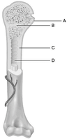
Key Terms:
Compact Bone: Dense, strong bone tissue.
Epiphyseal Line: Remnant of growth plate.
Yellow Marrow: Fat storage.
Epiphysis: End of the bone.
Step-by-Step Guidance
Examine the diagram and locate each labeled structure.
Recall the function of each part (e.g., where marrow is stored).
Match the description to the correct label (e.g., compact bone, epiphyseal line).
Review the role of each structure in bone growth and function.
Try solving on your own before revealing the answer!
Final Answer:
8) A: Compact bone, 9) B: Epiphyseal line, 10) C: Yellow marrow, 11) D: Epiphysis.
Q7. Using Figure 5.1, match the following skin structures and functions.
Background
Topic: Integumentary System Anatomy
This question tests your ability to identify skin structures and their functions.
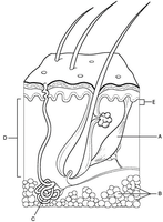
Key Terms:
Dermal Ridges: Produce fingerprints.
Hypodermis: Shock absorption, fat storage.
Arrector Pili Muscle: Causes hair to stand up.
Sudoriferous Gland: Sweat gland.
Dense Irregular Connective Tissue: Provides strength.
Step-by-Step Guidance
Examine the diagram and locate each labeled structure.
Recall the function of each part (e.g., which structure produces sweat).
Match the description to the correct label (e.g., dermal ridges, hypodermis).
Review the role of each structure in skin physiology.
Try solving on your own before revealing the answer!
Final Answer:
1) D: Dermal ridges, 2) C: Hypodermis, 3) A: Arrector pili muscle, 4) B: Sudoriferous gland, 5) E: Dense irregular connective tissue, 6) C: Hypodermis, 7) D: Capillary loops.
