 뒤로
Comprehensive Study Guide: Bones, Joints, and Muscles of the Human Body
스터디 가이드 - 스마트 노트
 자료에 맞춘 맞춤형 노트, 핵심 정의, 예시, 맥락을 확장해 제공합니다.
Bones & Skeletal Tissue
Osseous Tissue Types
The human skeleton is composed of two main types of osseous (bone) tissue: compact bone and spongy bone. Each type has distinct structural and functional characteristics.
Compact Bone: Located deep to the periosteum, composed of osteons (Haversian systems), provides strength and support.
Spongy Bone: Lacks Haversian canals, composed of trabeculae, found deep to compact bone, contains red bone marrow, resists compression.
Bone tissue functions: Mineral storage, movement, protection, red blood cell formation, triglyceride storage, and support.
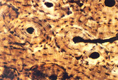
Bone Cells and Structure
Osteocytes: Mature bone cells found in lacunae.
Osteons: Functional subunits of compact bone, consisting of concentric lamellae surrounding a central canal.
Canaliculi: Tiny channels connecting lacunae, allowing communication and nutrient exchange.
Bone Shapes
Short bones: Carpals
Long bones: Humerus
Irregular bones: Vertebrae
Sesamoid bones: Patella
Flat bones: Scapula
Bone Anatomy
Periosteum: Outer layer of compact bone, anchored by Sharpey's fibers.
Lamellae Types:
Circumferential: Run parallel to bone surface.
Concentric: Circular layers around central canal.
Interstitial: Fill gaps between osteons.
Central (Haversian) Canal: Contains blood vessels and nerves, runs longitudinally.
Perforating (Volkmann's) Canal: Runs perpendicular, connects central canals.
The Skeletal System
Axial Skeleton
The axial skeleton forms the central supporting axis of the body and includes the skull, vertebrae, sacrum, sternum, ribs, and hyoid bone.
Accessory structures: Hyaline cartilage, fibrocartilage, elastic cartilage.
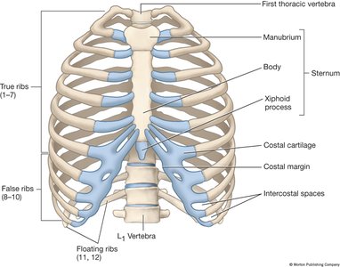
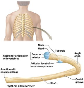
Rib Cage Anatomy
True ribs (1-7): Directly attached to sternum via costal cartilage.
False ribs (8-10): Indirectly attached to sternum.
Floating ribs (11-12): Not attached to sternum.
Sternum: Bone to which costal cartilage attaches.
Appendicular Skeleton
The appendicular skeleton consists of the limbs and girdles, excluding the vertebral column, trunk, and skull.
Scapula: Right or left determined by anatomical landmarks.
Clavicle: Medial (sternal) and lateral (acromial) ends.
Humerus: Right or left determined by deltoid tuberosity and anatomical neck.
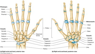
Wrist and Hand Anatomy
Carpals: Scaphoid, lunate, triquetrum, pisiform, hamate, capitate, trapezoid, trapezium.
Metacarpals: Head, body, base.
Phalanges: Distal, middle, proximal.
Pollex: Thumb region.
Joints
Classification of Joints
Joints are classified by structure and function.
Structural types:
Fibrous: Skull sutures, gomphosis, syndesmosis.
Cartilaginous: Synchondrosis (costochondral), symphysis (intervertebral discs).
Synovial: Shoulder, knee.
Functional types:
Synarthrosis: Immovable
Amphiarthrosis: Slightly movable
Diarthrosis: Freely movable
Synovial Joint Movements
Non-axial: Plane joints (intercarpals)
Uni-axial: Pivot (atlantoaxial), hinge (elbow)
Bi-axial: Condylar (metacarpophalangeal), saddle (thumb)
Multi-axial: Ball and socket (shoulder, hip)
Muscle Tissue
Muscle Tissue Types and Structure
Muscle tissue is classified as skeletal, cardiac, or smooth, each with unique features.
Skeletal muscle: Multinucleated, striated, voluntary, contains fascicles.
Cardiac muscle: Branched, involuntary, contains intercalated discs.
Smooth muscle: Involuntary, lacks striations and intercalated discs.
Muscle Cell Anatomy
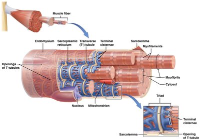
Sarcolemma: Plasma membrane of muscle cells.
Sarcoplasm: Cytoplasm of muscle cells.
Myofibrils: Rod-like structures containing contractile elements.
Sarcoplasmic reticulum: Stores and releases calcium ions.
T-tubules: Carry action potentials deep into muscle fibers.
Striations: Alternating dark (A bands, myosin) and light (I bands, actin) bands.
Sarcomere: Smallest functional unit of muscle contraction.
Muscle Contraction Process
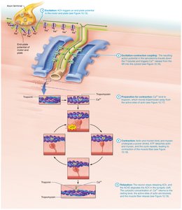
Excitation: Acetylcholine (ACh) triggers action potential in motor end plate.
Excitation-Contraction Coupling: Action potential travels down T-tubules, triggers calcium release from sarcoplasmic reticulum.
Preparation for Contraction: Calcium binds to troponin, tropomyosin moves away from actin's active site.
Contraction: Actin and myosin bind, myosin powerstroke, ATP detaches actin and myosin, cycle repeats.
Relaxation: ACh degraded, calcium returns to normal, actin and myosin sites blocked, muscle relaxes.
Muscle Attachments and Naming
Origin: Stationary bony attachment.
Insertion: Mobile bony attachment.
Belly: Region between origin and insertion.
Innervation: Nerve stimulating muscle contraction.
Muscle actions: Flexor, extensor, abductor, adductor.
Shapes: Deltoid (triangle), trapezius (trapezoid).
Sizes: Maximus, longus, brevis.
Fiber directions: Rectus (parallel), transversus, oblique.
Origins: Biceps, triceps, quadriceps.
Fascicle Arrangements
Circular: Sphincters
Convergent: Single tendon
Parallel: Strap-like or fusiform
Pennate: Unipennate, bipennate, multipennate (diagonal fibers)
Muscles of the Body
Neck Muscles
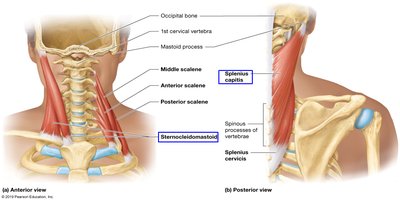
Sternocleidomastoid: Flexes and rotates the head.
Splenius capitis: Extends and rotates the head.
Thoracic Muscles
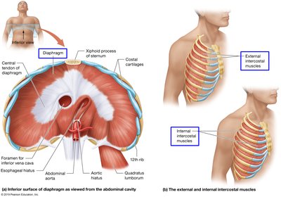
Diaphragm: Separates thoracic and abdominopelvic cavities, contracts for inhalation.
External intercostals: Aid inhalation.
Internal intercostals: Aid exhalation.
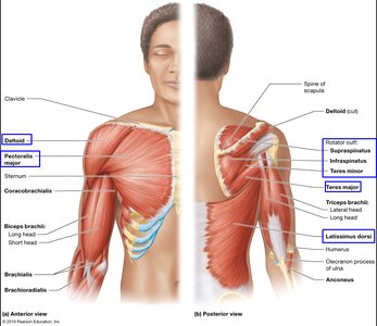
Shoulder and Arm Muscles
Deltoid: Abducts arm.
Pectoralis major: Flexes, adducts, and rotates arm.
Rotator cuff: Supraspinatus, infraspinatus, teres minor, stabilize shoulder.
Latissimus dorsi: Extends, adducts, and rotates arm.
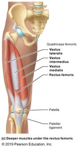
Thigh Muscles
Quadriceps femoris: Vastus lateralis, vastus intermedius, vastus medialis, rectus femoris; extend knee.
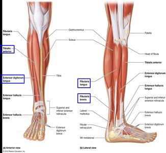
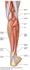
Lower Leg Muscles
Anterior: Tibialis anterior, extensor digitorum longus.
Lateral: Fibularis longus, fibularis brevis.
Posterior: Gastrocnemius, soleus, flexor digitorum longus, tibialis posterior.
Summary Table: Bone and Muscle Features
Feature | Bone | Muscle |
|---|---|---|
Cell Type | Osteocyte | Muscle fiber |
Functional Unit | Osteon | Sarcomere |
Matrix | Lamellae | Myofibrils |
Attachment | Periosteum | Epimysium |
Movement | Joint articulation | Contraction |
Key Equations
Muscle contraction:
Bone strength:
Additional info:
Some muscle and bone names are inferred based on standard anatomical knowledge.
Tables and images are recreated and expanded for clarity and completeness.
