 뒤로
뒤로Comprehensive Study Guide: Joints (Articulations) in Human Anatomy
스터디 가이드 - 스마트 노트
 자료에 맞춘 맞춤형 노트, 핵심 정의, 예시, 맥락을 확장해 제공합니다.
Joints: An Overview
Definition and Importance
Joints, also known as articulations, are the sites where two or more bones meet. They play a crucial role in providing mobility to the skeleton and enabling resistance to crushing, tearing, and other forces. The scientific study of joints is called arthrology (from the Greek root arthro, meaning joint).
Classification of Joints
Functional Classification
Joints are classified by the degree of movement they allow:
Synarthroses: Immovable joints; common in the axial skeleton.
Amphiarthroses: Slightly movable joints; also common in the axial skeleton.
Diarthroses: Freely movable joints; prevalent in the appendicular skeleton (all synovial joints).
Structural Classification
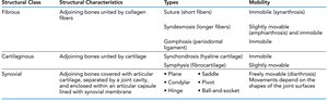
Structural classification is based on the material binding the bones and the presence or absence of a joint cavity. The main types are:
Fibrous Joints: Bones joined by fibrous connective tissue.
Cartilaginous Joints: Bones joined by cartilage.
Synovial Joints: Bones covered with articular cartilage and enclosed within a joint capsule lined with synovial membrane.
Structural Class | Structural Characteristics | Types | Mobility |
|---|---|---|---|
Fibrous | Adjoining bones united by collagen fibers | Suture (short fibers), Syndesmosis (longer fibers), Gomphosis (periodontal ligament) | Immobile (synarthrosis), Slightly movable (amphiarthrosis), Immobile |
Cartilaginous | Adjoining bones united by cartilage | Synchondrosis (hyaline cartilage), Symphysis (fibrocartilage) | Immobile, Slightly movable |
Synovial | Adjoining bones covered with articular cartilage, separated by a joint cavity, and enclosed within an articular capsule lined with synovial membrane | Plane, Hinge, Pivot, Condylar, Saddle, Ball-and-socket | Freely movable (diarthrosis); movements depend on shapes of the joint surfaces |
Fibrous Joints
General Characteristics
Fibrous joints are connected by dense regular connective tissue and lack a joint cavity. Most are immovable or only slightly movable.
Sutures: Occur only between bones of the skull; tightly bound by minimal fibrous tissue. Allow bone growth during childhood; later ossify to form synostoses.
Syndesmoses: Bones connected exclusively by ligaments. Movement depends on fiber length (e.g., tibiofibular joint—little movement; interosseous membrane between radius and ulna—more movement).
Gomphoses: Peg-in-socket joint; e.g., tooth in a socket, connected by the periodontal ligament.
Cartilaginous Joints
General Characteristics
Cartilaginous joints unite bones with cartilage and lack a joint cavity. They are not highly movable.
Synchondroses: Bones united by hyaline cartilage; immovable (e.g., epiphyseal plates, joint between first rib and manubrium).
Symphyses: Bones united by fibrocartilage; slightly movable, providing strength and flexibility (e.g., intervertebral discs, pubic symphysis). Hyaline cartilage is present as articular cartilage to reduce friction.
Synovial Joints
General Structure
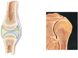
Synovial joints are the most movable type and all are diarthroses. Most joints in the limbs are synovial. Each contains a fluid-filled joint cavity.
Articular cartilage: Covers bone ends with hyaline cartilage, absorbing compression and preventing bone damage.
Joint (articular) cavity: Unique to synovial joints; holds synovial fluid.
Articular capsule: Encloses the joint cavity with a two-layered capsule (fibrous layer for strength, synovial membrane for fluid production).
Synovial fluid: Viscous, blood filtrate containing glycoproteins; lubricates and nourishes cartilage via "weeping lubrication".
Reinforcing ligaments: Strengthen the joint; may be extracapsular or intracapsular.
Sensory nerves: Detect pain and monitor capsule stretch (proprioception).
Blood supply: Richly supplied; capillary beds produce synovial fluid.
Articular Discs
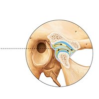
Some synovial joints contain articular discs (e.g., temporomandibular and knee joints) to improve fit, distribute load, minimize wear, and allow multiple movements.
Bursae and Tendon Sheaths
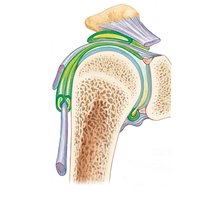
Bursa: Flattened fibrous sac lined by synovial membrane; reduces friction where body elements rub together.
Tendon sheath: Elongated bursa wrapping around a tendon.
Movements Allowed by Synovial Joints
Types of Movement
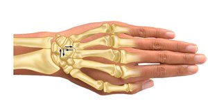
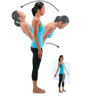
Gliding: One bone slides across another; occurs in carpals, tarsals, and vertebral articular processes.
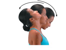
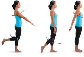
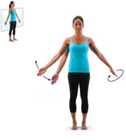
Angular Movements: Change the angle between bones. Includes flexion, extension, hyperextension, abduction, adduction, and circumduction.
Rotation: Bone turns around its long axis; occurs at atlas/axis, hip, shoulder, and vertebral column.
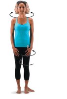
Special Movements
Elevation/Depression: Lifting or lowering a body part.
Protraction/Retraction: Moving a body part anteriorly or posteriorly.
Supination/Pronation: Rotating the forearm so the palm faces up (supination) or down (pronation).
Opposition: Thumb touches the tips of other fingers.
Dorsiflexion/Plantar Flexion: Up-and-down movements of the foot.
Inversion/Eversion: Turning the sole of the foot medially or laterally.
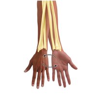
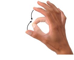
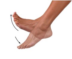
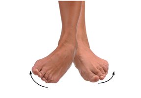
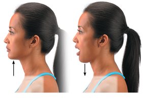
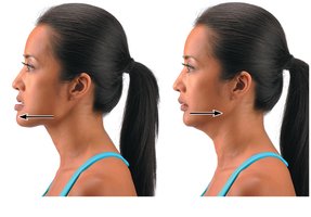
Synovial Joints: Functional and Structural Classification
Functional Classification
Nonaxial: Gliding movements only.
Uniaxial: Movement in one axis.
Biaxial: Movement in two axes.
Multiaxial: Movement in all three axes.
Synovial Joints Classified by Shape
Plane Joint: Flat articular surfaces; nonaxial gliding movements (e.g., intercarpal joints).
Hinge Joint: Cylindrical end fits into a trough; uniaxial movement (e.g., elbow, ankle).
Pivot Joint: Rounded bone turns around its long axis; uniaxial (e.g., proximal radioulnar joint).
Condylar (Ellipsoid) Joint: Oval surfaces; biaxial movement (e.g., metacarpophalangeal joints).
Saddle Joint: Each surface is concave and convex; biaxial (e.g., thumb carpometacarpal joint).
Ball-and-Socket Joint: Spherical head fits into socket; multiaxial (e.g., shoulder, hip).
Factors Influencing Stability of Synovial Joints
Articular Surfaces
The shape of articulating surfaces determines possible movements and, in some cases, provides stability (notably in the hip, elbow, and ankle).
Ligaments
Ligaments and capsules prevent excessive motion. The number and position of ligaments affect joint strength and stability.
Muscle Tone
Muscle tone stabilizes joints by keeping tension on tendons, especially important in the shoulder, knee, and foot arches.
Selected Synovial Joints
Temporomandibular Joint (TMJ)
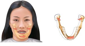
The TMJ is a modified hinge joint with articular surfaces covered by fibrocartilage and an articular disc dividing the cavity. It allows hingelike and gliding movements, as well as lateral excursion (side-to-side movement). 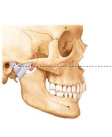
Sternoclavicular Joint
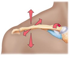
A saddle joint with an articular disc, surrounded by four ligaments. It allows multiple complex movements including elevation, depression, protraction, retraction, and rotation. 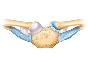
Shoulder (Glenohumeral) Joint
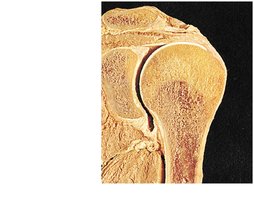
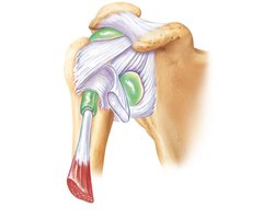
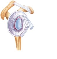
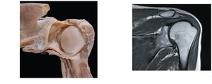
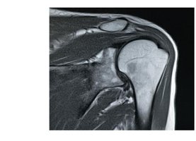
The most freely movable joint, but lacks stability. Stability is provided by the rotator cuff muscles and ligaments. The glenoid cavity is deepened by the glenoid labrum. 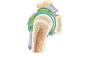
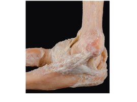
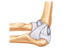
Elbow Joint
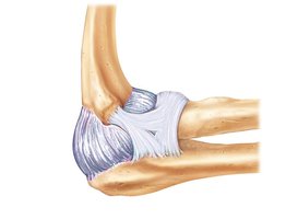
A stable hinge joint allowing only flexion and extension. Stability is provided by the articulation of the humerus and ulna, as well as tendons of biceps and triceps. 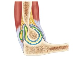
Wrist Joint
Composed of radiocarpal and intercarpal joints, stabilized by numerous ligaments. Allows flexion, extension, adduction, abduction, circumduction, and gliding movements. 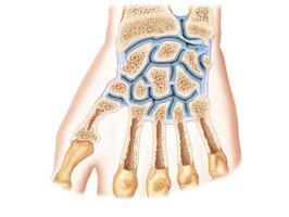
Hip Joint
A ball-and-socket joint with movements in all axes, limited by ligaments and the acetabulum. Stability is mainly from the acetabulum and capsular ligaments. 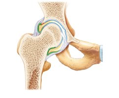
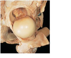
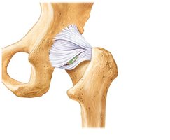
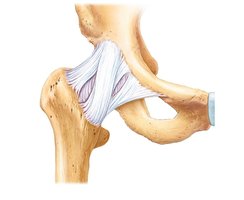
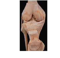
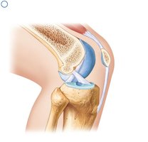
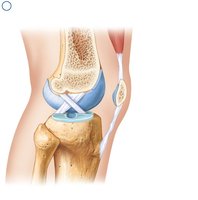
Knee Joint
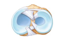
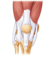
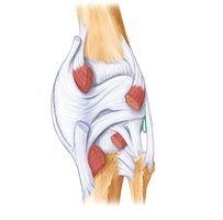
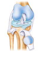
The largest and most complex joint, acting primarily as a hinge. Contains two fibrocartilage menisci and several ligaments for stability. 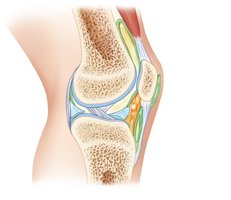
Ankle Joint
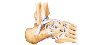
A hinge joint between the tibia, fibula, and talus. Allows dorsiflexion and plantar flexion. Stability is provided by medial (deltoid) and lateral ligaments, as well as tibiofibular ligaments. 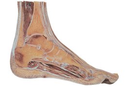
Summary Table: Joint Classes
Structural Class | Structural Characteristics | Types | Mobility |
|---|---|---|---|
Fibrous | Adjoining bones united by collagen fibers | Suture, Syndesmosis, Gomphosis | Immobile or slightly movable |
Cartilaginous | Adjoining bones united by cartilage | Synchondrosis, Symphysis | Immobile or slightly movable |
Synovial | Articular cartilage, joint cavity, capsule | Plane, Hinge, Pivot, Condylar, Saddle, Ball-and-socket | Freely movable |
Key Terms:
Arthrology: Scientific study of joints.
Synarthrosis: Immovable joint.
Amphiarthrosis: Slightly movable joint.
Diarthrosis: Freely movable joint.
Articular cartilage: Hyaline cartilage covering bone ends in synovial joints.
Meniscus: Fibrocartilage pad in the knee joint.
Bursa: Synovial membrane-lined sac reducing friction.
Example: The knee joint is a compound synovial joint, stabilized by ligaments, menisci, and muscle tone, allowing flexion, extension, and limited rotation. Additional info: This guide expands on brief points with academic context, definitions, and examples for clarity and completeness.
