 뒤로
Comprehensive Study Guide: Skeletal and Muscular Systems for Anatomy & Physiology Lab Practical
스터디 가이드 - 스마트 노트
 자료에 맞춘 맞춤형 노트, 핵심 정의, 예시, 맥락을 확장해 제공합니다.
Skeletal System
Bone Histology
The microscopic structure of bone reveals specialized features that support its function in the body. Understanding bone histology is essential for identifying bone types and their roles.
Osteon (Haversian System): The fundamental functional unit of compact bone, consisting of concentric lamellae arranged around a central canal containing blood vessels and nerves.
Spongy Bone vs. Compact Bone:
Compact bone is dense and forms the outer layer of bones, providing strength for weight-bearing.
Spongy bone (cancellous bone) is found at the ends of long bones and inside flat bones, containing trabeculae that support marrow.
Location in Long Bones: Compact bone forms the diaphysis (shaft), while spongy bone is found in the epiphyses (ends).
Connective Tissue Coverings:
Periosteum: Dense connective tissue covering the external surface of bones; contains osteoblasts and osteoclasts.
Endosteum: Thin membrane lining the internal bone surfaces, including the medullary cavity.
Skeletal Gross Anatomy
The human skeleton is divided into axial and appendicular components, each with distinct bones and functions.
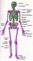
Axial Skeleton: Includes the skull, vertebral column, ribs, and sternum. Provides central support and protects vital organs.
Appendicular Skeleton: Comprises the limbs and girdles (pectoral and pelvic), facilitating movement and interaction with the environment.
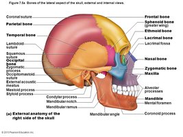
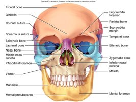
Bone Identification: Key bones of the skull (e.g., parietal, temporal, frontal, occipital, sphenoid, ethmoid, mandible, maxilla, zygomatic, nasal, lacrimal) and their landmarks (e.g., sutures, foramina, processes) are essential for anatomical orientation.
Shape and Function: Bones are classified by shape (long, short, flat, irregular, sesamoid) and serve functions such as support, protection, movement, mineral storage, and hematopoiesis.
Major Bones of the Axial Skeleton
Skull: Parietal, temporal, frontal, occipital, sphenoid, ethmoid, mandible, maxilla, zygomatic, nasal, lacrimal
Vertebral Column: 7 cervical, 12 thoracic, 5 lumbar vertebrae, sacrum (5 fused), coccyx
Thoracic Cage: Sternum (manubrium, body, xiphoid process), ribs
Hyoid bone
Major Bones of the Appendicular Skeleton
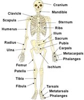
Pectoral Girdle: Clavicle, scapula
Upper Limb: Humerus, radius, ulna, carpals, metacarpals, phalanges
Pelvic Girdle: Ilium, ischium, pubis, sacrum, coccyx, pubic symphysis, acetabulum, iliac crest
Lower Limb: Femur, patella, tibia, fibula, tarsals, metatarsals, phalanges
Distinguishing Male and Female Skeletons
Pelvic Differences: Female pelvis is wider and shallower for childbirth; male pelvis is narrower and deeper.
Other Features: Males generally have more prominent bone markings due to larger muscle attachments.
Spinal Curvatures and Disorders
Normal Curves: Cervical and lumbar (lordotic), thoracic and sacral (kyphotic)
Abnormal Curvatures:
Kyphosis: Excessive posterior curvature, usually in thoracic region
Lordosis: Excessive anterior curvature, usually in lumbar region
Scoliosis: Lateral curvature of the spine
Articulations and Movements
Joints (articulations) connect bones and allow for movement. They are classified by structure and function.
Structural Classification:
Fibrous Joints: Bones joined by dense connective tissue (e.g., sutures of the skull)
Cartilaginous Joints: Bones joined by cartilage (e.g., intervertebral discs)
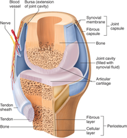
Synovial Joints: Bones separated by a joint cavity filled with synovial fluid; most movable type
Functional Classification: Synarthrosis (immovable), amphiarthrosis (slightly movable), diarthrosis (freely movable)
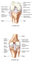
Synovial Joint Structure: Articular cartilage, joint (synovial) cavity, articular capsule (fibrous and synovial layers), synovial fluid, ligaments, nerves, and blood vessels
Types of Synovial Joints and Movements
Types: Plane, hinge, pivot, condyloid, saddle, ball-and-socket
Movements: Flexion/extension, abduction/adduction, circumduction, rotation, pronation/supination, dorsiflexion/plantar flexion, elevation/depression, protraction/retraction, opposition/reposition, eversion/inversion
Muscular System
Muscle Histology
Muscle tissue is specialized for contraction and is classified into three types based on structure and function.
Skeletal Muscle: Striated, voluntary, multinucleated; attached to bones for movement
Cardiac Muscle: Striated, involuntary, branched, with intercalated discs; found in the heart
Smooth Muscle: Non-striated, involuntary, spindle-shaped; found in walls of hollow organs
Sarcomere Structure
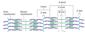
Sarcomere: The functional contractile unit of skeletal muscle, defined by Z-disks
Key Components: Actin (thin) and myosin (thick) myofilaments, A band, I band, H zone, M line, Z disk
Muscle Organization
Muscle Fiber: Single muscle cell
Endomysium: Connective tissue surrounding each muscle fiber
Perimysium: Surrounds bundles of fibers (fascicles)
Epimysium: Surrounds the entire muscle
Fascia: Connective tissue that separates muscles from each other
Muscle Physiology
Muscle contraction is influenced by electrical and mechanical factors.
Effect of Voltage: Increasing stimulus voltage recruits more muscle fibers, increasing tension (up to a maximum).
Frequency of Stimulus: Higher frequency leads to summation and tetanus (sustained contraction).
Muscle Length: Optimal length allows maximal cross-bridge formation and tension generation.
Additional info: Refer to PhysioEx simulations for practical demonstrations of these principles.
Gross Muscle Anatomy
Understanding muscle anatomy involves identifying origins, insertions, shapes, and connective tissue structures.
Origin: The fixed attachment point of a muscle
Insertion: The movable attachment point
Muscle Shapes: Circular (orbicularis), pennate (rectus femoris), parallel (sartorius), convergent (pectoralis major), fusiform (biceps brachii)
Tendon: Dense connective tissue attaching muscle to bone
Aponeurosis: Broad, flat tendon (e.g., epicranial aponeurosis)
Muscle Actions: Flexion, extension, abduction, adduction, rotation, etc.
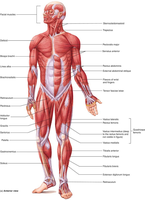
Muscle Labeling and Actions
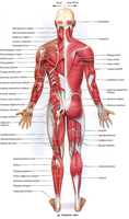
Be able to identify major muscles on diagrams and models
Know the primary action of each muscle (e.g., gracilis adducts the thigh)
Summary Table: Comparison of Bone and Muscle Tissue Types
Feature | Compact Bone | Spongy Bone | Skeletal Muscle | Cardiac Muscle | Smooth Muscle |
|---|---|---|---|---|---|
Location | Shaft of long bones | Ends of long bones, inside flat bones | Attached to skeleton | Heart | Walls of hollow organs |
Structure | Osteons, dense | Trabeculae, porous | Striated, multinucleated | Striated, branched, intercalated discs | Non-striated, spindle-shaped |
Control | Involuntary | Involuntary | Voluntary | Involuntary | Involuntary |
