 뒤로
Comprehensive Study Guide: Skeletal, Cardiac, and Smooth Muscles & Muscle Naming in Anatomy and Physiology
스터디 가이드 - 스마트 노트
 자료에 맞춘 맞춤형 노트, 핵심 정의, 예시, 맥락을 확장해 제공합니다.
Skeletal Muscle System
Levers in the Human Body
Muscles and bones interact as levers to produce movement. Levers are classified based on the relative positions of the fulcrum, load, and effort:
First Class Lever: The fulcrum is positioned between the load and the effort. Example: nodding the head.
Second Class Lever: The load is between the fulcrum and the effort. Example: standing on tiptoe.
Third Class Lever: The effort is applied between the fulcrum and the load. This is the most common lever in the body, such as the action of the hamstrings.
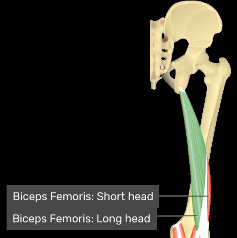
Hamstrings are part of a third class lever, the most common in the body. Quadriceps are the prime movers of knee extension, while hamstrings are antagonists and flex the knee.
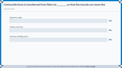
Origin: The fixed attachment of a muscle.
Insertion: The movable attachment that moves toward the origin during contraction.
Cardiac Muscle Tissue
Structure and Function
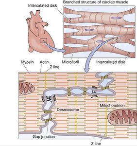
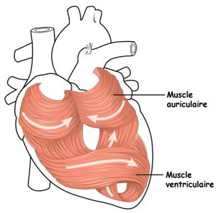
Cardiac muscle is found only in the heart. Its fibers are branched, have a single nucleus, and contain short, wide T-tubules. Cardiac muscle lacks triads and cisternae, and relies on aerobic metabolism for energy.
Intercalated Discs: Specialized cell connections that allow cardiac muscle to beat in rhythm as a syncytium, stabilize adjacent cells, and maintain the 3D structure of the tissue.
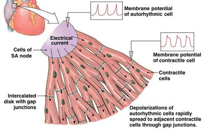
Autorhythmic Cells: Pacemaker cells can initiate contraction without neural stimulation; the nervous system alters the pace.
Contractile Cells: Have longer contraction and relaxation phases, allowing for filling and pumping periods. The sarcolemma prevents wave summation and tetanus.
Smooth Muscle Tissue
Structure and Types
Smooth muscle is found in the walls of hollow organs. Cells are elongated, spindle-shaped, and contain a single nucleus. Smooth muscle is non-striated (lacks sarcomeres), involuntary, and does not have tendons or aponeuroses.
Unitary (Visceral) Fibers: Slow contractions, involuntary control, myogenic with neuronal input, not connected to motor neurons, cells depolarize based on local conditions and hormones.
Multiunit Fibers: Fast contractions, involuntary control, neurogenic, neuron ends on single cell creating many small motor units, each cell connected to more than one neuron.
Actions of Dorsal and Ventral Muscles
Muscle Actions Based on Position
In the anatomical position:
Dorsal Muscles: Extend, elevate, protract, and abduct.
Ventral Muscles: Flex, depress, retract, and adduct.
Muscles that Move the Vertebral Column
Erector Spinae and Deep Muscles
The erector spinae group extends and laterally flexes the spine. It consists of three main muscles:
Spinalis: Most medial, extends and laterally flexes spine.
Longissimus: Intermediate, extends, laterally flexes, and rotates head and spine.
Iliocostalis: Most lateral, extends and laterally flexes spine.
Deep muscles include semispinalis (extends and rotates spine), rotatores (extend and rotate spine), and quadratus lumborum (lateral flexion and stabilization).
Muscles that Move the Thigh and Leg
Hip and Knee Movements
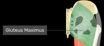
Major movements at the hip include flexion (iliopsoas), extension (gluteus maximus), abduction (gluteus medius and minimus), adduction (gracilis, adductor longus, adductor magnus), and rotation (tensor fascia latae, sartorius).
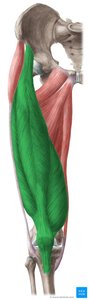
Knee flexion is performed by the hamstrings (biceps femoris, semimembranosus, semitendinosus), while extension is performed by the quadriceps femoris (rectus femoris, vastus lateralis, vastus intermedius, vastus medialis).
Muscles that Move the Foot
Ankle Movements
Key movements include dorsiflexion (tibialis anterior), plantarflexion (gastrocnemius, soleus, fibularis longus and brevis), inversion (tibialis posterior), and eversion (fibularis longus and brevis).
Muscles of the Abdomen
Abdominal Muscle Actions
Abdominal muscles include external oblique, internal oblique, transversus abdominus, and rectus abdominus. They rotate the trunk, compress the abdomen, and flex the vertebral column.
Muscles that Move the Arm
Shoulder and Arm Movements
Major movements include flexion (pectoralis major, deltoid, biceps brachii), extension (latissimus dorsi, posterior deltoid, teres major), adduction (latissimus dorsi, teres major), abduction (deltoid, supraspinatus), rotation (infraspinatus, teres minor, subscapularis, teres major).
Muscles that Move the Wrist
Wrist Movements
Prime movers for wrist flexion include flexor carpi ulnaris and flexor carpi radialis; for extension, extensor carpi ulnaris and extensor carpi radialis; for adduction, flexor carpi ulnaris and extensor carpi ulnaris; for abduction, flexor carpi radialis and extensor carpi radialis.
Muscles Used in Respiration
Respiratory Muscles
Key muscles for inhalation include diaphragm, external intercostals, sternocleidomastoid, scalenes, serratus anterior, and pectoralis minor. Exhalation involves internal intercostals and abdominal muscles.
Muscles of Facial Expression and Mastication
Facial Muscles
Muscles include occipitofrontalis (raises eyebrows), orbicularis oculi (closes eyelids), orbicularis oris (puckers lips), zygomaticus major (pulls lips laterally and superiorly), buccinator (compresses cheeks).
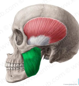
Muscles of Mastication
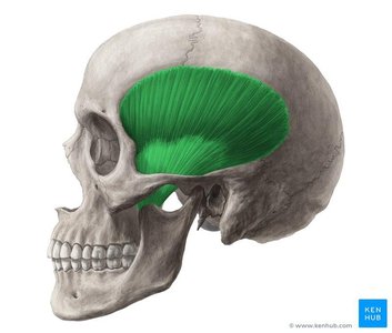
Masseter and temporalis elevate the mandible for chewing.
Muscles that Move the Head
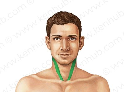
Sternocleidomastoid rotates and flexes the head; splenius capitis extends and flexes the head.
Muscles that Move the Shoulder Girdle
Pectoralis minor and serratus anterior stabilize and protract the scapula; rhomboid major and minor retract the scapula; trapezius retracts, rotates, depresses, and elevates the scapula; levator scapulae elevates the scapula.
Muscles that Move the Toes
Extensor hallicus longus and extensor digitorum longus extend the toes; flexor hallicus longus and flexor digitorum longus flex the toes.
Muscles that Move the Forearm
Biceps brachii and brachialis flex the forearm; triceps brachii extends the forearm; pronator teres and pronator quadratus pronate the forearm; supinator supinates the forearm.
Muscles that Move the Fingers
Flexor digitorum superficialis and profundus flex the digits; flexor pollicis longus flexes the thumb; extensor digitorum and extensor pollicis brevis extend the digits and thumb; abductor pollicis longus and brevis abduct the thumb; adductor pollicis adducts the thumb; dorsal and palmar interossei adduct digits; opponens pollicis brings thumb and digit 5 together.
