 뒤로
Comprehensive Study Guide: The Skeletal System
스터디 가이드 - 스마트 노트
 자료에 맞춘 맞춤형 노트, 핵심 정의, 예시, 맥락을 확장해 제공합니다.
The Skeletal System
Overview and Functions
The skeletal system is a complex framework of bones and connective tissues that provides structural support, protection, and facilitates movement. It also serves as a reservoir for minerals and houses bone marrow for blood cell production.
Bone Tissue: Bone is a specialized connective tissue composed of cells (osteocytes, osteoblasts, osteoclasts) and a mineralized matrix.
Functions: Support, protection of internal organs, movement (with muscles), mineral storage (calcium, phosphate), and blood cell formation (hematopoiesis).
Divisions of the Skeletal System
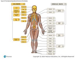
The human skeleton is divided into two main regions: the axial and appendicular skeletons.
Axial Skeleton: Includes the skull, vertebral column, and thoracic cage.
Appendicular Skeleton: Includes the limbs, pectoral girdle, and pelvic girdle.
Total Bones: The adult human skeleton contains 206 bones.
Classification of Bones
Bones are classified based on their shape and function.
Long Bones: e.g., femur, humerus
Short Bones: e.g., carpals, tarsals
Flat Bones: e.g., skull, sternum
Irregular Bones: e.g., vertebrae, pelvis
Surface Markings and Parts of a Long Bone
Surface markings on bones serve as attachment points for muscles, tendons, and ligaments, and as passageways for nerves and blood vessels. The structure of a typical long bone includes several distinct regions.
Diaphysis: Shaft of the bone
Epiphysis: Ends of the bone
Metaphysis: Region between diaphysis and epiphysis
Medullary Cavity: Central cavity containing bone marrow
Periosteum: Outer covering of bone
Endosteum: Inner lining of the bone
Microscopic Structure of Bone
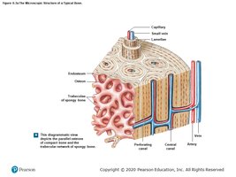
Bones are composed of compact and spongy tissue, each with unique structural features.
Compact Bone: Dense, forms the outer layer
Spongy Bone: Porous, found at the ends of long bones
Osteon: Structural unit of compact bone
Canaliculi: Small channels for nutrient and waste exchange
Bone Cells and Growth
Bone maintenance and growth are regulated by three main cell types and processes.
Osteocytes: Mature bone cells, maintain bone matrix
Osteoblasts: Bone-forming cells
Osteoclasts: Bone-resorbing cells
Appositional Growth: Increase in bone diameter by adding new layers to the surface
Cartilage vs. Bone
Cartilage and bone are both connective tissues but differ in structure and function.
Cartilage: Flexible, avascular, found in joints, ear, nose
Bone: Rigid, vascular, provides structural support
Skull Bones and Sutures
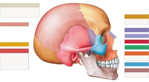
The adult skull consists of several bones joined by sutures, which are immovable joints.
Major Bones: Frontal, parietal, occipital, temporal, sphenoid, ethmoid, zygomatic, maxilla, mandible, nasal, lacrimal
Sutures: Coronal (frontal-parietal), squamous (temporal-parietal), lambdoid (parietal-occipital)
Vertebral Column and Spinal Curves
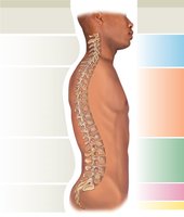
The vertebral column is divided into regions, each with distinct structural characteristics and functions. Spinal curves are classified as primary (present at birth) and secondary (develop after birth).
Cervical (7 vertebrae): Secondary curve, develops as infant balances head
Thoracic (12 vertebrae): Primary curve, present at birth
Lumbar (5 vertebrae): Secondary curve, develops with standing
Sacral and Coccygeal: Primary curves, present at birth
Function: Protects spinal cord, supports head and trunk, allows flexibility
Classification of Ribs
Ribs are classified based on their articulation with the sternum.
True Ribs: Directly attached to sternum (1-7)
False Ribs: Indirectly attached or not attached (8-12)
Floating Ribs: Not attached to sternum (11-12)
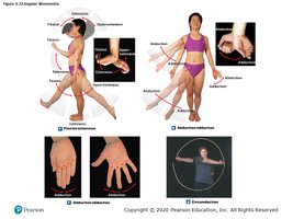
Movements of the Body
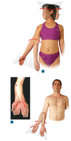
Body movements are produced by the interaction of bones, muscles, and joints. Specific terms describe these movements.
Flexion/Extension: Decrease/increase angle between bones
Abduction/Adduction: Move limb away/toward midline
Circumduction: Circular movement
Rotation: Movement around longitudinal axis
Joints: Classification and Structure
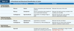
Joints are classified by their structure and function. The main types are fibrous, cartilaginous, and synovial joints.
Synarthrosis: Immovable joints (e.g., sutures)
Amphiarthrosis: Slightly movable joints (e.g., symphysis)
Diarthrosis: Freely movable joints (e.g., synovial joints)
Functional Category | Structural Category and Type | Description | Example |
|---|---|---|---|
Synarthrosis (No Movement) | Fibrous - Suture | Fibrous connections plus interlocked surfaces | Between bones of the skull |
Synarthrosis (No Movement) | Fibrous - Gomphosis | Fibrous connection plus insertion in bony socket | Between teeth and jaw sockets |
Synarthrosis (No Movement) | Cartilaginous - Synchondrosis | Rigid cartilage bridge or plate | Between first pair of ribs and sternum |
Amphiarthrosis (Little Movement) | Fibrous - Syndesmosis | Ligamentous connection | Between tibia and fibula |
Amphiarthrosis (Little Movement) | Cartilaginous - Symphysis | Connection by fibrocartilage pad | Between adjacent vertebrae or pubic bones |
Diarthrosis (Free Movement) | Synovial | Complex joint bounded by joint capsule and containing synovial fluid | Shoulder, hip, knee |
Synovial Joints: Structure and Types
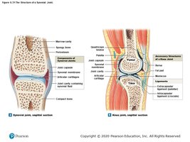
Synovial joints are characterized by a joint capsule, synovial fluid, and accessory structures. They allow free movement and are classified by the shape of their articulating surfaces.
Plane (Gliding) Joint: Flat surfaces, slight movement
Hinge Joint: Angular movement in one plane
Condylar (Ellipsoidal) Joint: Angular movement in two planes
Saddle Joint: Angular movement and circumduction
Pivot Joint: Rotation in one plane
Ball-and-Socket Joint: Angular, rotational, and circumduction movement
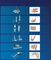
Parts of a Synovial Joint
Each synovial joint contains specific structures that contribute to its function.
Joint Capsule: Encloses the joint
Synovial Membrane: Produces synovial fluid
Articular Cartilage: Reduces friction
Ligaments: Stabilize the joint
Menisci: Pads for shock absorption
Summary Table: Types of Synovial Joints
Type | Movement | Example |
|---|---|---|
Plane (Gliding) | Multidirectional in a single plane | Carpals, tarsals |
Hinge | Angular in a single plane | Elbow, knee |
Condylar (Ellipsoidal) | Angular in two planes | Wrist, fingers |
Saddle | Angular in two planes, circumduction | Thumb |
Pivot | Rotation in a single plane | Atlas-axis, radius-ulna |
Ball-and-Socket | Angular, rotation, circumduction | Shoulder, hip |
Additional info:
Fractures are classified by their pattern and location; common types include transverse, oblique, spiral, and comminuted.
Surface markings include foramina (holes), processes (projections), and fossae (depressions).
Bone remodeling is a continuous process involving osteoblasts and osteoclasts.
