 뒤로
Comprehensive Study Notes on Joints (Articulations) in the Skeletal System
스터디 가이드 - 스마트 노트
 자료에 맞춘 맞춤형 노트, 핵심 정의, 예시, 맥락을 확장해 제공합니다.
Joints (Articulations) of the Skeletal System
Introduction to Joints
Joints, also known as articulations, are the sites where two bones meet. They serve two fundamental functions: providing mobility to the skeleton and holding the skeleton together. Joints are classified both structurally and functionally, based on their composition and the degree of movement they allow.
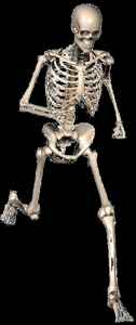
Structural Classification of Joints
Structural classification is based on the material binding the bones and the presence or absence of a joint cavity. There are three main structural types:
Fibrous Joints: Bones are joined by fibrous tissue; no joint cavity.
Cartilaginous Joints: Bones are joined by cartilage; no joint cavity.
Synovial Joints: Bones are separated by a fluid-filled joint cavity.
Fibrous Joints
Fibrous joints are united by dense connective tissue and allow little to no movement. Types include:
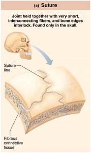
Sutures: Found only in the skull; short fibers interlock bone edges.
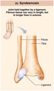
Syndesmoses: Bones connected by a ligament; allows slight movement. Example: distal tibia and fibula.
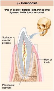
Gomphoses: Peg-in-socket joints; example: tooth in its socket.
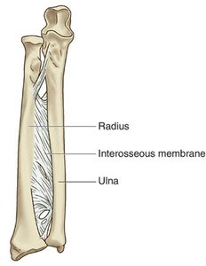
Interosseous Membrane: Connects radius and ulna in the forearm.
Cartilaginous Joints
Cartilaginous joints unite bones with cartilage and allow more movement than fibrous joints but less than synovial joints. Types include:
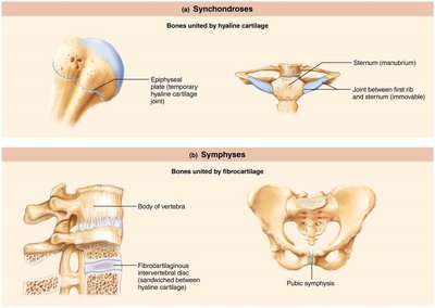
Synchondroses: Bones united by hyaline cartilage; mostly immovable. Example: epiphyseal plate, joint between first rib and sternum.
Symphyses: Bones united by fibrocartilage; slightly movable. Example: intervertebral discs, pubic symphysis.
Synovial Joints
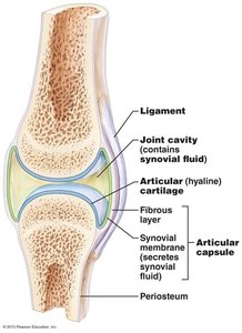
Synovial joints are the most movable type of joint and are characterized by a joint cavity filled with synovial fluid. They are found in most limb joints.
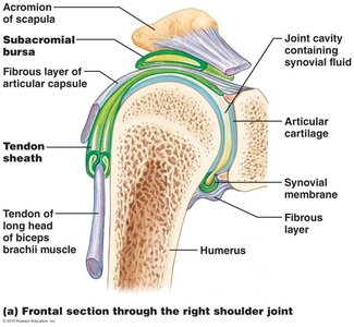
Joint (synovial) cavity: Space containing synovial fluid.
Articular cartilage: Hyaline cartilage covering bone surfaces.
Articular capsule: Two layers—outer fibrous layer and inner synovial membrane.
Synovial fluid: Lubricates and nourishes the joint.
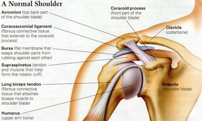
Reinforcing ligaments: Strengthen the joint.
Nerves and blood vessels: Supply the synovial membrane and provide filtrate for synovial fluid.
Structures Associated with Synovial Joints
Bursae: Sacs lined with synovial membrane that reduce friction where ligaments, muscles, skin, tendons, or bones rub together.
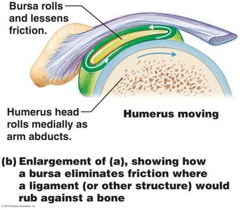
Tendon Sheaths: Elongated bursae wrapped around tendons subjected to friction.
Stabilizing Factors at Synovial Joints
Stability of synovial joints is maintained by:
Shapes of articular surfaces
Ligament number and location
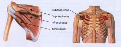
Muscle tendons that cross the joint (muscle tone keeps tendons taut)
Ligaments of Major Joints
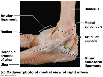
Elbow Joint
Ulnar Collateral Ligament: Provides medial stability to the elbow.
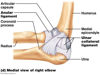
Cadaver View: Shows anatomical structures of the elbow.
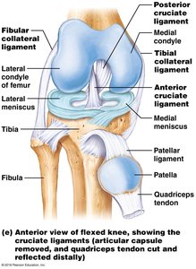
Knee Joint
Anterior Cruciate Ligament (ACL): Prevents anterior displacement of the tibia.
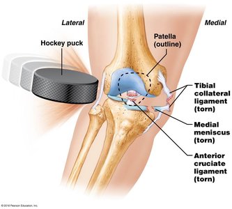
ACL Injury: Common sports injury affecting knee stability.
Types of Synovial Joints
Synovial joints are classified by the shape of their articular surfaces and the type of movement they allow:
Plane Joint: Flat surfaces, gliding movement. Example: intercarpal joints.
Hinge Joint: Cylinder and trough, uniaxial movement (flexion/extension). Example: elbow.
Pivot Joint: Rounded bone fits into a sleeve, uniaxial rotation. Example: proximal radioulnar joint.
Condylar Joint: Oval surfaces, biaxial movement (flexion/extension, abduction/adduction). Example: knuckle joints.
Saddle Joint: Concave and convex surfaces, biaxial movement. Example: thumb joint.
Ball-and-Socket Joint: Spherical head fits into a cup, multiaxial movement. Example: shoulder and hip joints.
Movements Allowed by Synovial Joints
Synovial joints allow a wide range of movements, classified as gliding, angular, and rotational. Special movements include supination, pronation, dorsiflexion, plantar flexion, inversion, eversion, protraction, retraction, elevation, depression, and opposition.
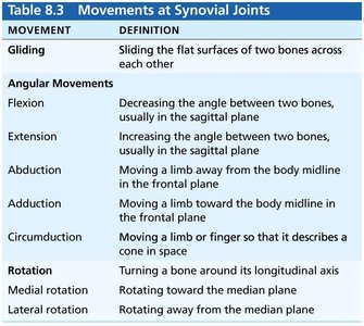
Movement | Definition |
|---|---|
Gliding | Sliding the flat surfaces of two bones across each other |
Flexion | Decreasing the angle between two bones, usually in the sagittal plane |
Extension | Increasing the angle between two bones, usually in the sagittal plane |
Abduction | Moving a limb away from the body midline in the frontal plane |
Adduction | Moving a limb toward the body midline in the frontal plane |
Circumduction | Moving a limb or finger so that it describes a cone in space |
Rotation | Turning a bone around its longitudinal axis |
Medial rotation | Rotating toward the median plane |
Lateral rotation | Rotating away from the median plane |
Joint Examples and Clinical Relevance
Major synovial joints include the temporomandibular joint (TMJ), elbow, knee, hip, and shoulder. These joints are highly mobile but also at risk for injury due to their complexity and range of motion. Stability is provided by ligaments, tendons, and muscle tone.
Example: The shoulder joint is a ball-and-socket joint, allowing multiaxial movement but is prone to dislocation due to its shallow socket.
Example: The knee joint is a hinge joint, stabilized by multiple ligaments including the ACL, which is commonly injured in sports.
Additional info: Synovial fluid is produced by the synovial membrane and acts as a lubricant and nutrient source for articular cartilage. Bursae and tendon sheaths are specialized structures that further reduce friction and facilitate smooth movement.
