 뒤로
Comprehensive Study Notes on Joints (Articulations) in Human Anatomy & Physiology
스터디 가이드 - 스마트 노트
 자료에 맞춘 맞춤형 노트, 핵심 정의, 예시, 맥락을 확장해 제공합니다.
Joints: Structure, Classification, and Function
Introduction to Joints
Joints, also known as articulations, are anatomical structures where two or more bones meet. They are essential for providing mobility to the skeleton while maintaining its integrity. The study of joints is called arthrology, and the study of movement is known as kinesiology.
Point of Contact: Joints can occur between two bones, between cartilage and bone, or between teeth and bones.
Function: Joints hold bones together and permit movement, making body motion possible.

Classification of Joints
Structural and Functional Classification
Joints are classified based on their structure and function:
Structural Classification: Based on the presence or absence of a synovial cavity and the type of connective tissue binding the bones.
Functional Classification: Based on the degree of movement permitted.
Structural Type | Synovial Cavity | Connecting Tissue |
|---|---|---|
Fibrous | Absent | Fibrous connective tissue |
Cartilaginous | Absent | Cartilage (hyaline or fibrocartilage) |
Synovial | Present | Articular capsule and ligaments |
Functional Type | Degree of Movement |
|---|---|
Synarthrosis | Immovable |
Amphiarthrosis | Slightly movable |
Diarthrosis | Freely movable |
Fibrous Joints
General Features
Fibrous joints lack a synovial cavity and are held together by dense fibrous connective tissue. They allow little or no movement, functioning as synarthroses or amphiarthroses.
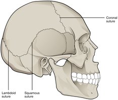
Sutures: Immovable joints found between bones of the skull.
Syndesmoses: Bones connected by a ligament; slightly movable.
Gomphoses: Peg-in-socket joints, such as teeth in their sockets; immovable.
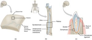
Sutures
Sutures are thin layers of dense fibrous connective tissue that unite the bones of the skull. They are immovable (synarthrosis) and may fuse completely in adults, forming a synostosis.
Syndesmoses
Syndesmoses are fibrous joints where bones are united by a ligament, allowing slight movement (amphiarthrosis). Examples include the anterior tibiofibular joint and the interosseous membrane between the radius and ulna.
Gomphoses
Gomphoses are specialized fibrous joints where a ligament holds a cone-shaped peg (tooth) in a bony socket (alveolus). These are immovable joints (synarthrosis).
Cartilaginous Joints
General Features
Cartilaginous joints lack a synovial cavity and allow little or no movement. The bones are tightly connected by either hyaline cartilage or fibrocartilage.
Synchondroses: Bones joined by hyaline cartilage; immovable (synarthrosis).
Symphyses: Bones joined by fibrocartilage; slightly movable (amphiarthrosis).
Synchondroses
In synchondroses, the connecting material is hyaline cartilage. These joints are immovable and include the epiphyseal plates of growing bones and the joints between the ribs and sternum.
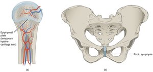
Symphyses
Symphyses are cartilaginous joints where the bones are united by fibrocartilage, allowing slight movement. Examples include the intervertebral discs and the pubic symphysis.
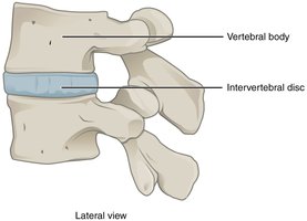
Synovial Joints
General Features
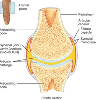
Synovial joints are characterized by the presence of a synovial cavity that separates the articulating bones. These joints are freely movable (diarthroses) and are the most common type of joint in the body.
Articular cartilage: Covers the ends of bones, reducing friction and absorbing shock.
Articular capsule: Surrounds the joint, consisting of an outer fibrous capsule and an inner synovial membrane.
Synovial fluid: Secreted by the synovial membrane, lubricates the joint and nourishes the cartilage.
Ligaments: Thickenings in the fibrous capsule that provide support and limit movement.
Types of Synovial Joints
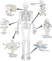
There are six main types of synovial joints, each allowing different types of movement:
Type | Movement | Example |
|---|---|---|
Pivot | Rotation | Atlantoaxial joint (C1-C2) |
Hinge | Flexion/Extension | Elbow, knee |
Saddle | Variety, including opposition | Thumb (carpometacarpal joint) |
Plane | Gliding | Intertarsal joints |
Condyloid | Flexion/Extension, Abduction/Adduction | Wrist |
Ball-and-Socket | Multiaxial movement | Shoulder, hip |
Accessory Structures of Synovial Joints
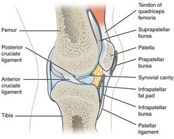
Bursae: Fluid-filled sacs that reduce friction between moving structures such as skin, muscle, or tendon and bone.
Tendon Sheaths: Tubular bursae that surround tendons, especially in the wrist and ankle.
Movements at Synovial Joints
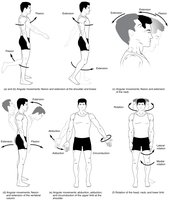
Synovial joints allow a wide range of movements, including:
Flexion/Extension: Decreasing/increasing the angle between bones (e.g., bending and straightening the elbow).
Abduction/Adduction: Moving a limb away from/toward the midline of the body.
Circumduction: Circular movement combining flexion, extension, abduction, and adduction.
Rotation: Turning a bone around its own axis.
Special Movements
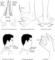
Supination/Pronation: Rotational movement of the forearm.
Dorsiflexion/Plantar Flexion: Movements at the ankle joint.
Eversion/Inversion: Movements of the sole of the foot.
Protraction/Retraction: Anterior/posterior movement of a body part (e.g., mandible).
Elevation/Depression: Lifting/lowering a body part (e.g., mandible).
Opposition/Reposition: Movement of the thumb to touch other fingers and back.
Selected Synovial Joints of the Body
Atlantoaxial Joint
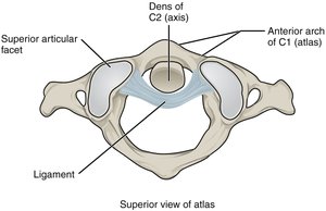
The atlantoaxial joint is a pivot joint between the dens of the axis (C2) and the anterior arch of the atlas (C1), allowing rotation of the head.
Temporomandibular Joint (TMJ)
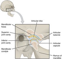
The TMJ is the articulation between the temporal bone and the mandible, containing an articular disc. It allows both hinge and gliding movements.
Glenohumeral (Shoulder) Joint
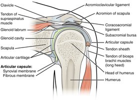
The shoulder joint is a ball-and-socket joint with the greatest range of motion. It is supported by ligaments and the rotator cuff muscles.
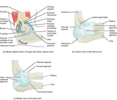
Elbow Joint
The elbow is a hinge joint allowing flexion and extension. It is stabilized by the ulnar and radial collateral ligaments and the annular ligament.
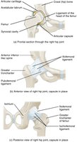
Hip Joint
The hip is a ball-and-socket joint providing stability and a wide range of motion. Ligaments tighten when standing, securing the femoral head in the acetabulum.
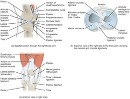
Knee Joint
The knee is the largest joint in the body, functioning as a hinge joint. It is supported by collateral and cruciate ligaments, and the menisci provide cushioning.
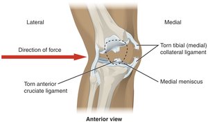
Knee Injuries
A strong lateral blow to the knee can cause a sequence of injuries: tearing of the tibial collateral ligament, damage to the medial meniscus, and rupture of the anterior cruciate ligament (ACL).
Ankle Joint
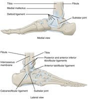
The talocrural (ankle) joint is a uniaxial hinge joint allowing dorsiflexion and plantar flexion. Ligaments support the joint and resist excessive movements.
Clinical Consideration: Osteoarthritis
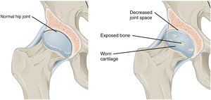
Osteoarthritis is a degenerative joint disease resulting from aging or prolonged wear and tear. It leads to erosion of articular cartilage, inflammation, joint stiffness, and pain.
Summary Table: Joint Types and Examples
Joint Type | Structural Classification | Functional Classification | Example |
|---|---|---|---|
Suture | Fibrous | Synarthrosis | Skull bones |
Syndesmosis | Fibrous | Amphiarthrosis | Distal tibiofibular joint |
Gomphosis | Fibrous | Synarthrosis | Teeth in sockets |
Synchondrosis | Cartilaginous | Synarthrosis | Epiphyseal plate |
Symphysis | Cartilaginous | Amphiarthrosis | Pubic symphysis |
Synovial | Synovial | Diarthrosis | Shoulder, knee |
