 뒤로
뒤로Excitation-Contraction Coupling in Skeletal Muscle
스터디 가이드 - 스마트 노트
 자료에 맞춘 맞춤형 노트, 핵심 정의, 예시, 맥락을 확장해 제공합니다.
Excitation-Contraction Coupling in Skeletal Muscle
Overview of Excitation-Contraction Coupling (E-CC)
Excitation-Contraction Coupling (E-CC) is the physiological process by which an electrical stimulus (action potential) is converted into a mechanical response (muscle contraction). This process is fundamental to skeletal muscle function and involves a series of tightly regulated steps at the neuromuscular junction and within the muscle fiber.
Key Point 1: E-CC links the nervous system to muscle contraction via neurotransmitter release and ion channel activation.
Key Point 2: Calcium ions play a central role in initiating and terminating muscle contraction.
Example: A muscle twitch is a single contraction-relaxation cycle triggered by a single action potential.
Sequence of Events: Acetylcholine Release to Muscle Twitch
The process begins at the neuromuscular junction (NMJ), where the somatic motor neuron releases acetylcholine (ACh), leading to muscle fiber activation and contraction.
Step 1: Somatic motor neuron releases acetylcholine (ACh) at the NMJ.
Step 2: ACh binds to receptors on the sarcolemma (muscle cell membrane), causing ion channels to open.
Step 3: Sodium ions enter the cell, potassium ions exit, resulting in depolarization and formation of the end-plate potential (EPP).
Step 4: EPP triggers a muscle action potential that propagates along the sarcolemma and into the T-tubules.
Step 5: The action potential activates L-type calcium channel dihydropyridine (DHP) receptors on the T-tubule membrane.
Step 6: DHP receptors mechanically interact with ryanodine receptors (RyR) on the sarcoplasmic reticulum, causing calcium release.
Step 7: Calcium binds to troponin, initiating contraction by allowing actin-myosin cross-bridge formation.
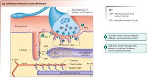
Role of DHP and Ryanodine Receptors in E-CC
DHP and Ryanodine receptors are critical for the transmission of the action potential from the T-tubule to the sarcoplasmic reticulum, facilitating calcium release.
DHP Receptor: A voltage-sensitive L-type calcium channel located in the T-tubule membrane; acts as a sensor for the action potential.
Ryanodine Receptor (RyR): A calcium release channel in the sarcoplasmic reticulum; named after the plant toxin ryanodine, which binds to and modulates its activity.
Clinical Relevance: Calcium channel blockers (targeting DHP receptors) are used to lower blood pressure; ryanodine is a potent insecticide.
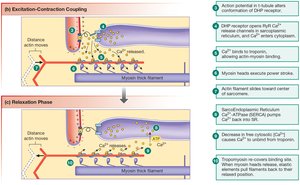
Calcium's Role in Muscle Contraction and Relaxation
Calcium ions are essential for both the initiation and termination of muscle contraction. Their movement is tightly regulated by membrane channels and pumps.
Contraction: Calcium released from the sarcoplasmic reticulum binds to troponin, causing tropomyosin to move and exposing actin binding sites for myosin.
Relaxation: Calcium is actively pumped back into the sarcoplasmic reticulum by the SERCA ATPase, allowing tropomyosin to cover actin binding sites and ending contraction.
Equation:
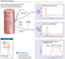
Timing of Excitation-Contraction Coupling: Muscle Twitch and Latent Period
A muscle twitch consists of a single contraction-relaxation cycle. The latent period is the brief delay between the action potential and the onset of muscle tension, reflecting the time required for calcium release and binding to troponin.
Latent Period: The interval between the muscle action potential and the beginning of muscle tension development.
Significance: Represents the time needed for E-CC processes, including calcium release and cross-bridge formation.
Graphical Representation: Muscle twitch graphs show the action potential, latent period, contraction, and relaxation phases.
Summary Table: Key Steps in Excitation-Contraction Coupling
This table summarizes the main events and their physiological significance in E-CC.
Step | Event | Physiological Significance |
|---|---|---|
1 | ACh release at NMJ | Initiates muscle activation |
2 | Action potential in sarcolemma | Triggers downstream events |
3 | DHP receptor activation | Detects voltage change |
4 | RyR opens, Ca++ release | Ca++ enters cytosol |
5 | Ca++ binds troponin | Initiates contraction |
6 | SERCA pumps Ca++ back | Causes relaxation |
Learning Objectives
Describe the sequence of events between acetylcholine release at the NMJ and a muscle twitch.
Define DHP and Ryanodine receptors and their roles in E-CC.
Explain the role of Ca++ in muscle contraction and relaxation.
Define the latent period in excitation-contraction coupling.
