 뒤로
Functional Organization of the Muscular System: Structure, Classification, and Mechanics
스터디 가이드 - 스마트 노트
 자료에 맞춘 맞춤형 노트, 핵심 정의, 예시, 맥락을 확장해 제공합니다.
Functional Organization of the Muscular System
Overview of the Muscular System
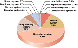
The muscular system is a major contributor to body mass, accounting for approximately half of the total body weight. It consists of about 700 skeletal muscles, which vary widely in shape, size, and function. Muscle performance is determined by factors such as muscle size, shape, fascicle arrangement, and skeletal attachments.
Muscle size and shape: Influence force production and range of motion.
Fascicle arrangement: Determines the muscle's functional capabilities.
Skeletal attachments: Affect leverage and movement efficiency.
Divisions of Skeletal Muscles
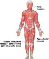
Skeletal muscles are divided into two main groups based on their location and function:
Axial muscles: Position and stabilize the head, neck, and trunk; assist in breathing.
Appendicular muscles: Move and stabilize limbs and their girdles.
Tendons: Connect skeletal muscles to bones, transmitting muscle force to bones.
Fascicle Organization and Muscle Architecture
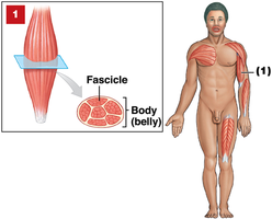
Parallel Fascicle Arrangement
In parallel muscles, fascicles run parallel to the long axis of the muscle, often resulting in a prominent muscle belly. These muscles allow for relatively large shortening and excursion, making them well-suited for movements requiring a wide range of motion. Example: biceps brachii.
Convergent Fascicle Arrangement
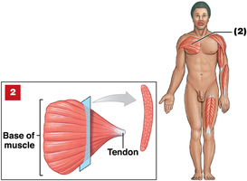
Convergent muscles have a broad origin that converges toward a common tendon or insertion. This arrangement allows different regions of the muscle to contribute to different lines of pull, providing versatility in movement. Example: pectoralis major.
Pennate Fascicle Arrangement
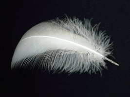
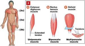
Pennate muscles have fascicles that attach obliquely to a tendon, forming an angle resembling a feather. This architecture packs more fibers into a given muscle volume than parallel muscles, typically resulting in greater force capacity but less excursion.
Unipennate: Fibers on one side of a tendon (e.g., extensor digitorum).
Bipennate: Fibers on both sides of a tendon (e.g., rectus femoris).
Multipennate: Multiple internal tendon branches and featherlike fascicles (e.g., deltoid).
Circular Fascicle Arrangement (Sphincters)
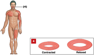
Circular muscles or sphincters have fascicles arranged in concentric circles, encircling a duct, tube, or opening. Contraction decreases the diameter of the opening, effectively constricting it. Example: orbicularis oris.
Summary Table: Fascicle Arrangements
Arrangement | Structure | Function | Example |
|---|---|---|---|
Parallel | Fascicles along long axis | Large excursion | Biceps brachii |
Convergent | Broad base, narrow attachment | Versatile pull | Pectoralis major |
Pennate | Angled fibers | Force optimized | Deltoid, rectus femoris |
Circular | Rings | Constriction | Orbicularis oris |
Levers and Movement
Lever Systems in the Body
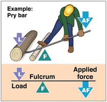
A lever is a rigid structure (often a bone) that rotates around a fulcrum (pivot point, usually a joint). Muscles provide the effort (force), while the load is the resistance. Lever geometry influences force, speed, and distance of movement.
First-class lever: Fulcrum is between applied force and load (e.g., head extension at the atlanto-occipital joint).
Second-class lever: Load is between fulcrum and applied force (e.g., rising onto the balls of the feet).
Third-class lever: Applied force is between fulcrum and load; most common in the body, favoring speed and range of motion.
Muscle Attachments and Actions
Origin, Insertion, and Action
Muscles attach to bones or connective tissues at two points:
Origin: Fixed end of a skeletal muscle, typically proximal.
Insertion: Movable end, typically distal.
Action: Specific movement produced by the muscle.
Agonists, Synergists, and Antagonists
Muscles work in groups to produce and control movement:
Agonist (prime mover): Muscle primarily responsible for movement (e.g., biceps brachii for elbow flexion).
Synergist: Assists the agonist, providing additional pull or stabilizing origin (e.g., brachioradialis).
Fixator: Synergist that prevents movement at another joint.
Antagonist: Opposes the action of the agonist (e.g., triceps brachii for elbow flexion).
Muscle Naming and Terminology
Muscle Names: Clues to Structure and Function
Muscle names often provide information about their location, position, direction, structure, and action. Understanding these terms aids in identification and memorization.
Location: Brachii (arm), femoris (thigh), abdominis (abdomen), capitis (head), carpi (wrist), oris (mouth).
Direction/Position: Rectus (straight), oblique (diagonal), transversus (across), medialis (toward midline), lateralis (away from midline), superficialis (near surface), profundus (deep).
Structure: Biceps (two heads), triceps (three heads), quadriceps (four heads), deltoid (triangular), orbicularis (circular), major/minor, maximus/minimus, longus/brevis.
Action: Flexor/extensor (bend/straighten), abductor/adductor (move away/toward midline), levator/depressor (raise/lower), pronator/supinator (rotate forearm).
Axial and Appendicular Muscle Classification
Axial Muscles
Arise on the axial skeleton and constitute about 60% of skeletal muscles. They position the head and spinal column and move the rib cage, assisting in breathing.
Appendicular Muscles
Make up the remaining 40% of skeletal muscles. They stabilize or move the appendicular skeleton (limbs and girdles).
Anterior and Posterior Muscle Identification
Axial examples: Temporalis, rectus abdominis, external oblique (anterior); posterior head/neck and trunk muscles (posterior).
Appendicular examples: Deltoid, pectoralis major, biceps brachii, rectus femoris (anterior); trapezius, latissimus dorsi, gluteus maximus, gastrocnemius (posterior).
Review and Application
Key Review Questions
Why can a pennate muscle produce more tension than a parallel muscle of similar size?
How are first-, second-, and third-class levers distinguished?
Which class describes the head balancing at the neck?
Decode muscle names: What can you infer from rectus abdominis, orbicularis oris, triceps brachii, extensor digitorum?
Summary Table: Muscle Terminology
Term | Meaning | Example |
|---|---|---|
Brachii | Arm | Biceps brachii |
Rectus | Straight | Rectus abdominis |
Orbicularis | Circular | Orbicularis oris |
Triceps | Three heads | Triceps brachii |
Extensor | Straightening movement | Extensor digitorum |
Additional info:
Pennate muscles produce more tension due to a greater number of fibers packed into the same volume.
Lever classes are distinguished by the relative positions of fulcrum, applied force, and load.
Muscle names can be decoded to reveal their action, location, and structure.
