 뒤로
Fundamentals of the Nervous System and Nervous Tissue: Study Guide
스터디 가이드 - 스마트 노트
 자료에 맞춘 맞춤형 노트, 핵심 정의, 예시, 맥락을 확장해 제공합니다.
Fundamentals of the Nervous System and Nervous Tissue
Characteristics and Functions of the Nervous System
The nervous system is a complex network responsible for controlling and coordinating bodily functions. It enables rapid communication between different parts of the body and is essential for sensation, integration, and response.
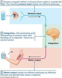
Sensory Input: The nervous system detects internal and external stimuli through sensory receptors. This information is transmitted via afferent pathways to the central nervous system (CNS).
Integration: The CNS processes and interprets sensory input, deciding on an appropriate response. This occurs in the brain or spinal cord.
Motor Output: The nervous system sends signals via efferent pathways to effectors (muscles and glands), causing a response.
Reflex Arc: A pathway that allows for rapid, automatic responses to stimuli.
Major Divisions of the Nervous System
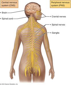
The nervous system is divided into two main structural and functional components: the central nervous system (CNS) and the peripheral nervous system (PNS).
Central Nervous System (CNS): Consists of the brain and spinal cord. It acts as the integrative and control center.
Peripheral Nervous System (PNS): Composed of cranial nerves, spinal nerves, and ganglia. It connects the CNS to the rest of the body.
General Organization of the Nervous System
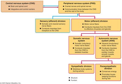
Sensory (Afferent) Division: Carries sensory information from receptors to the CNS.
Motor (Efferent) Division: Transmits commands from the CNS to effectors.
Somatic Nervous System: Controls voluntary movements (skeletal muscles).
Autonomic Nervous System (ANS): Regulates involuntary functions (smooth muscle, cardiac muscle, glands). Includes sympathetic and parasympathetic divisions.
Histology of Nervous Tissue
Nervous tissue consists of two main cell types: neurons and neuroglia (glial cells). Neurons are excitable cells that transmit electrical signals, while neuroglia provide support and protection.
Types of Neuroglia (Glial Cells)
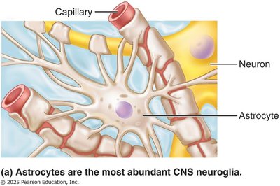
Astrocytes: Most abundant CNS glia; support neurons, regulate blood-brain barrier, and maintain extracellular environment.
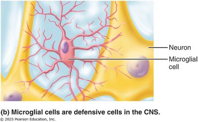
Microglial Cells: Defensive cells; act as macrophages to remove debris and pathogens.
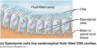
Ependymal Cells: Line cerebrospinal fluid-filled cavities; help circulate CSF.
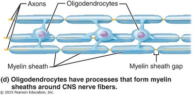
Oligodendrocytes: Form myelin sheaths in the CNS; myelinate multiple axons.
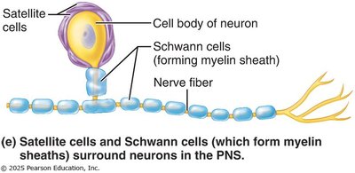
Satellite Cells: Surround neuron cell bodies in the PNS; regulate environment.
Schwann Cells: Form myelin sheaths in the PNS; myelinate one axon segment each.
Neurons: Structure and Function
Neurons are the structural and functional units of the nervous system. They are specialized for communication and possess unique properties.
Excitability: Ability to respond to stimuli and generate electrical signals.
Membrane Potential: Electrical charge difference across the cell membrane.
Action Potentials: Rapid electrical impulses transmitted along the neuron.
Longevity: Neurons can live for a lifetime.
Amitotic: Most neurons do not divide after development.
High Metabolic Rate: Require continuous supply of oxygen and glucose.
Major Structures of a Typical Neuron
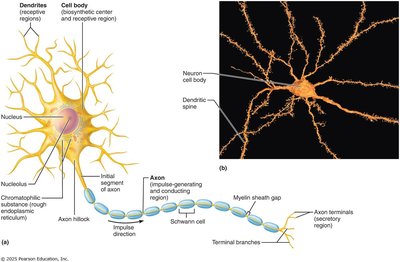
Cell Body (Soma): Contains nucleus and organelles; site of metabolic activity.
Dendrites: Receive input signals from other neurons.
Axon: Conducts nerve impulses away from the cell body.
Axon Hillock: Initiates action potentials.
Axon Terminals: Release neurotransmitters to communicate with other cells.
Neuron Groupings
Tracts: Bundles of axons in the CNS.
Nerves: Bundles of axons in the PNS.
Nuclei: Clusters of cell bodies in the CNS.
Ganglia: Clusters of cell bodies in the PNS.
Axonal Transport
Anterograde Transport: Movement from cell body to axon terminals (e.g., neurotransmitters).
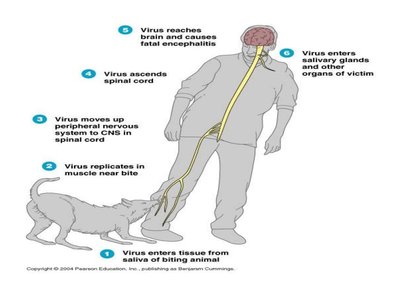
Retrograde Transport: Movement from axon terminals to cell body (e.g., recycled materials, viruses).
Functional Types of Neurons
Neurons are classified based on their function:
Sensory Neurons: Carry information from sensory receptors to the CNS.
Interneurons: Located within the CNS; process and integrate information.
Motor Neurons: Transmit commands from the CNS to effectors (muscles/glands).
Myelination and Myelin Sheath
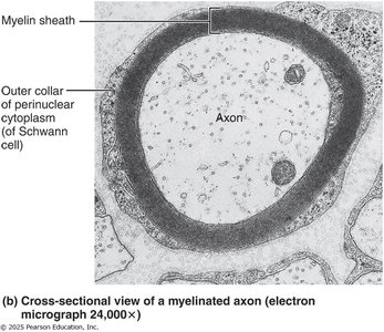
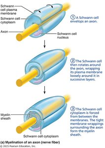
Myelin is a lipid-rich insulating material that surrounds axons, increasing the speed of nerve impulse conduction. Myelinated axons conduct impulses faster than unmyelinated axons.
Oligodendrocytes (CNS): Myelinate multiple axons.
Schwann Cells (PNS): Myelinate one axon segment each.
Nodes of Ranvier: Gaps between myelin segments; enable saltatory conduction (impulse jumps from node to node).
Comparison of Oligodendrocytes and Schwann Cells
Cell Type | Location | Function |
|---|---|---|
Oligodendrocyte | CNS | Myelinates multiple axons |
Schwann Cell | PNS | Myelinates one axon segment |
Disorders Associated with Axonal Transport
Rabies: Virus travels retrograde along axons to the CNS.
Herpes Simplex Virus (HSV): Uses axonal transport to reach the CNS.
Polio: Infects motor neurons, leading to paralysis.
Summary Table: Types of Glial Cells
Cell Type | Location | Function |
|---|---|---|
Astrocyte | CNS | Support, regulate environment |
Microglial Cell | CNS | Defense, phagocytosis |
Ependymal Cell | CNS | Line CSF cavities, circulate CSF |
Oligodendrocyte | CNS | Myelinate axons |
Satellite Cell | PNS | Support neuron cell bodies |
Schwann Cell | PNS | Myelinate axons |
Key Equations and Concepts
Membrane Potential: The voltage difference across a neuron's plasma membrane.
Saltatory Conduction: Rapid transmission of nerve impulses along myelinated axons, jumping from node to node.
Example: In multiple sclerosis, myelin is damaged, leading to slower nerve impulse conduction and neurological symptoms.
Additional info: The notes above expand on brief slide points to provide a comprehensive, exam-ready summary of nervous system fundamentals, histology, neuron structure, and myelination.
