 뒤로
Fundamentals of the Nervous System and Nervous Tissue: Membrane Potentials and Action Potentials
스터디 가이드 - 스마트 노트
 자료에 맞춘 맞춤형 노트, 핵심 정의, 예시, 맥락을 확장해 제공합니다.
Membrane Potentials: Graded Potentials
Definition and Production of Graded Potentials
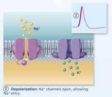
Graded potentials are local changes in membrane voltage that occur in response to a stimulus. They are essential for initiating action potentials and are produced when ion channels open, allowing ions to flow across the membrane. - Key Point 1: Graded potentials occur primarily in the dendrites and cell body of a neuron. - Key Point 2: The magnitude of a graded potential depends on the strength of the stimulus; stronger stimuli produce larger voltage changes. - Key Point 3: Graded potentials can be depolarizing (making the inside less negative) or hyperpolarizing (making the inside more negative). - Example: Sodium ions (Na+) entering the cell cause depolarization, while potassium ions (K+) leaving cause hyperpolarization. 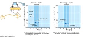
Locations and Stimuli Initiating Graded Potentials
Graded potentials are initiated at the dendrites or cell body by various stimuli, including neurotransmitter binding, mechanical deformation, or voltage changes. - Key Point 1: Chemically gated, voltage-gated, and mechanically gated ion channels are involved in generating graded potentials. - Key Point 2: Graded potentials decay with distance from the site of stimulation, making them local signals. 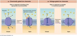
Membrane Potentials: Action Potentials
Definition and Triggering of Action Potentials
An action potential is a rapid, all-or-none electrical event that propagates along the axon, allowing long-distance communication within the nervous system. - Key Point 1: Action potentials are triggered when the membrane potential reaches a threshold value, typically around -55 mV. - Key Point 2: Action potentials do not fade with distance and are essential for transmitting signals from the axon hillock to the axon terminals. 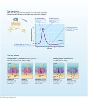
Phases of the Action Potential
The action potential consists of several distinct phases, each involving specific ion channel activity and ion movement.
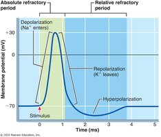
Phase | Membrane Potential | Ion Channel Activity | Ion Movement |
|---|---|---|---|
Resting State | -70 mV | All gated channels closed | Leaky Na+ in, K+ out |
Threshold | -55 mV | Voltage-gated Na+ channels open | Na+ influx |
Depolarization | +30 mV | Na+ channels open | Na+ influx |
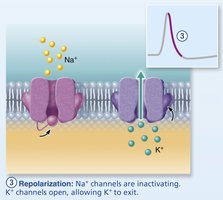
Repolarization | Falling toward -70 mV | Na+ channels inactivate, K+ channels open | K+ efflux |
Hyperpolarization | <-70 mV | Some K+ channels remain open | More K+ efflux |
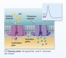
Comparison of Graded and Action Potentials
- Location: Graded potentials occur in dendrites and cell body; action potentials occur in axons. - Amplitude: Graded potentials vary in size; action potentials are all-or-none. - Duration: Graded potentials are brief and local; action potentials are longer and propagate. - Channel Types: Graded potentials use chemically, mechanically, and voltage-gated channels; action potentials use voltage-gated channels. - Function: Graded potentials initiate action potentials; action potentials transmit signals.
Resting Membrane Potential (RMP)
Establishment and Maintenance of RMP
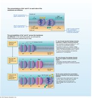
The resting membrane potential is the voltage difference across the plasma membrane when the neuron is not transmitting signals. - Key Point 1: RMP is typically -70 mV, with the inside of the neuron being more negative than the outside. - Key Point 2: The Na+/K+ ATPase pump maintains ion gradients by moving 3 Na+ out and 2 K+ in, using ATP. - Key Point 3: Permeability to K+ is especially important for maintaining RMP. 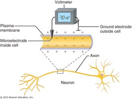
Action Potential Propagation and Coding for Stimulus Intensity
Propagation of Action Potentials
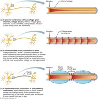
Action potentials begin at the axon hillock and propagate along the axon toward the terminals. - Key Point 1: Propagation is unidirectional due to refractory periods. - Key Point 2: Myelinated axons conduct action potentials faster via saltatory conduction (jumping between nodes of Ranvier). 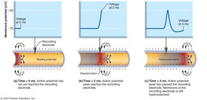
Coding for Stimulus Intensity
The central nervous system codes for stimulus intensity by the frequency of action potentials, not their amplitude. - Key Point 1: Stronger stimuli generate more frequent action potentials. - Key Point 2: Subthreshold stimuli do not generate action potentials. 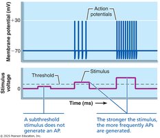
Refractory Periods
Absolute and Relative Refractory Periods
Refractory periods are phases during which a neuron cannot or is less likely to fire another action potential. - Absolute Refractory Period: No second action potential can occur, regardless of stimulus strength; voltage-gated Na+ channels are open or inactivating. - Relative Refractory Period: A second action potential can occur, but only with a stronger stimulus; membrane is hyperpolarized and some Na+ channels are recovering. 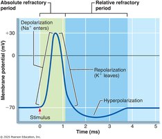
Abnormalities of Nervous Tissue
Multiple Sclerosis and Local Anesthesia
Disorders affecting nervous tissue can alter normal physiological function. - Multiple Sclerosis (MS): Demyelinating disorder that slows or blocks action potential conduction in the CNS. - Local Anesthesia: Blocks voltage-gated Na+ channels, preventing action potential generation and transmission. 
Key Ions in Membrane Potentials
Roles of Sodium, Potassium, Calcium, and Chloride
- Sodium (Na+): Major extracellular ion, influx causes depolarization. - Potassium (K+): Major intracellular ion, efflux causes repolarization and hyperpolarization. - Calcium (Ca2+): Important for neurotransmitter release. - Chloride (Cl-): Contributes to inhibitory postsynaptic potentials.
Summary Table: Ion Channel Types
Channel Type | Opening Mechanism | Function |
|---|---|---|
Leaky (non-gated) | Always open | Maintain RMP |
Voltage-gated | Voltage change | Action potentials |
Chemically gated | Ligand binding | Graded potentials |
Mechanically gated | Physical deformation | Graded potentials |
Equations and Formulas
Nernst Equation for Equilibrium Potential
The equilibrium potential for an ion can be calculated using the Nernst equation:
Goldman-Hodgkin-Katz Equation for Membrane Potential
The membrane potential considering multiple ions is given by:
Central and Peripheral Nervous System Overview
Structural Organization
The nervous system is divided into the central nervous system (CNS) and peripheral nervous system (PNS). - CNS: Includes the brain and spinal cord. - PNS: Includes cranial nerves, spinal nerves, and ganglia. 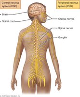
Additional info:
- Academic context was added to clarify the phases of action potentials, the role of ion channels, and the clinical relevance of disorders such as multiple sclerosis and local anesthesia. - Tables and equations were inferred and expanded for completeness and exam preparation.
