 뒤로
Fundamentals of the Nervous System and Nervous Tissue
스터디 가이드 - 스마트 노트
 자료에 맞춘 맞춤형 노트, 핵심 정의, 예시, 맥락을 확장해 제공합니다.
Fundamentals of the Nervous System and Nervous Tissue
Overview of the Nervous System
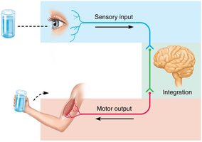
The nervous system is the master controlling and communicating system of the body. It uses electrical and chemical signals to coordinate rapid, specific, and immediate responses to internal and external stimuli.
Sensory Input: Information gathered by sensory receptors about internal and external changes.
Integration: Processing and interpretation of sensory input to determine an appropriate response.
Motor Output: Activation of effector organs (muscles and glands) to produce a response.
Structural and Functional Divisions of the Nervous System
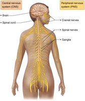
The nervous system is divided into two principal parts: the Central Nervous System (CNS) and the Peripheral Nervous System (PNS).
Central Nervous System (CNS): Consists of the brain and spinal cord. It is the integration and control center, interpreting sensory input and dictating motor output.
Peripheral Nervous System (PNS): Consists mainly of nerves that extend from the brain and spinal cord (cranial and spinal nerves). It serves as communication lines between the CNS and the rest of the body.
Functional Divisions of the PNS
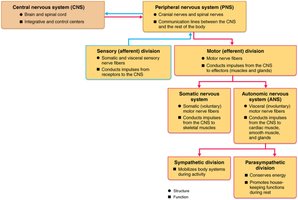
Sensory (Afferent) Division: Transmits sensory information to the CNS. Includes somatic sensory fibers (from skin, skeletal muscles, joints) and visceral sensory fibers (from visceral organs).
Motor (Efferent) Division: Transmits impulses from the CNS to effector organs. Subdivided into:
Somatic Nervous System: Conducts impulses to skeletal muscles; under voluntary control.
Autonomic Nervous System (ANS): Regulates smooth muscle, cardiac muscle, and glands; involuntary. Further divided into:
Sympathetic Division: Mobilizes body systems during activity.
Parasympathetic Division: Conserves energy and promotes housekeeping functions during rest.
Neuroglia and Neurons
Types of Neuroglia (Glial Cells)
Neuroglia are supporting cells in nervous tissue. They provide structural and metabolic support to neurons. There are distinct types in the CNS and PNS.
Neuroglia of the CNS
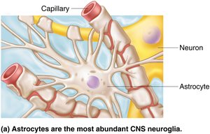
Astrocytes: Most abundant glial cells; support and brace neurons, aid in exchanges between capillaries and neurons, and participate in information processing in the brain.
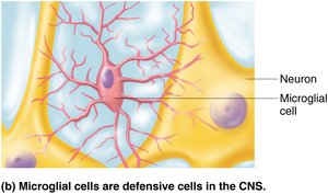
Microglial Cells: Small, defensive cells that monitor neuron health and can transform into phagocytes to remove debris and pathogens.
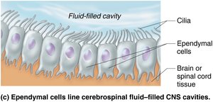
Ependymal Cells: Line cerebrospinal fluid-filled cavities in the CNS; their cilia help circulate cerebrospinal fluid (CSF).
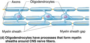
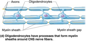
Oligodendrocytes: Branched cells that form insulating myelin sheaths around CNS nerve fibers.
Neuroglia of the PNS
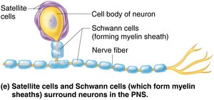
Satellite Cells: Surround neuron cell bodies in the PNS; function similarly to astrocytes in the CNS.
Schwann Cells (Neurolemmocytes): Surround all peripheral nerve fibers and form myelin sheaths in thicker fibers; vital for regeneration of damaged peripheral nerve fibers.
Neurons: Structure and Function
Neurons are the structural and functional units of the nervous system. They are highly specialized for impulse conduction and have unique properties:
Extreme longevity (can last a lifetime)
Amitotic (do not divide after maturity, with few exceptions)
High metabolic rate (require continuous oxygen and glucose)
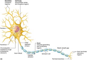
Consist of a cell body (soma) and one or more processes (dendrites and axon)
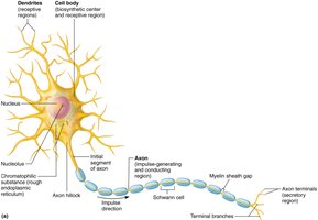
Neuron Cell Body (Soma)
Contains the nucleus and nucleolus; biosynthetic center of the neuron.
Most are located in the CNS (called nuclei); in the PNS, clusters are called ganglia.
Neuron Processes
Dendrites: Short, branched processes that receive input and convey it toward the cell body as graded potentials.
Axon: Single, long process that generates and transmits nerve impulses away from the cell body. Axons may branch (axon collaterals) and end in axon terminals (terminal boutons).
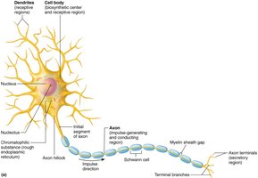
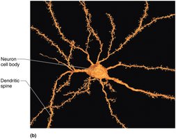
Axonal Transport
Anterograde: Movement away from the cell body (e.g., mitochondria, enzymes).
Retrograde: Movement toward the cell body (e.g., degraded organelles, viruses, toxins).
Myelin Sheath
The myelin sheath is a whitish, protein-lipid covering that insulates axons and increases the speed of nerve impulse transmission.
Myelinated fibers: Axons with a segmented myelin sheath; conduct impulses rapidly.
Nonmyelinated fibers: Axons without a myelin sheath; conduct impulses more slowly.
Myelination in the PNS
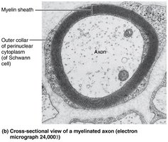
Formed by Schwann cells wrapping around the axon in a jelly roll fashion; one cell forms one segment of myelin sheath.
Myelin sheath gaps (nodes of Ranvier) are present between adjacent Schwann cells.
Nonmyelinated fibers are surrounded by Schwann cells but not wrapped in myelin.
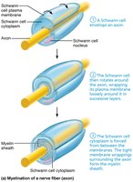
Myelination in the CNS
Formed by oligodendrocyte processes; each cell can myelinate multiple axons.
Myelin sheath gaps are present; no outer collar of perinuclear cytoplasm.
White matter: regions with dense collections of myelinated fibers (fiber tracts).
Gray matter: mostly neuron cell bodies and nonmyelinated fibers.
Classification of Neurons
Structural Classification
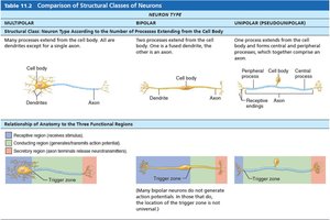
Neurons are classified by the number of processes extending from the cell body:
Type | Structure | Location/Example |
|---|---|---|
Multipolar | Three or more processes (1 axon, others dendrites) | Most common; major neuron type in CNS |
Bipolar | Two processes (1 axon, 1 dendrite) | Rare; retina, olfactory mucosa |
Unipolar (Pseudounipolar) | One T-like process (two axons) | Mainly in PNS; sensory neurons |
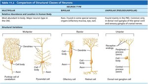
Functional Classification
Neurons are also classified by the direction in which the nerve impulse travels relative to the CNS:
Sensory (Afferent) Neurons: Transmit impulses from sensory receptors toward the CNS; almost all are unipolar, with cell bodies in ganglia in the PNS.
Motor (Efferent) Neurons: Carry impulses from the CNS to effectors (muscles/glands); multipolar, with most cell bodies in the CNS.
Interneurons (Association Neurons): Lie between motor and sensory neurons; shuttle signals through CNS pathways; most are entirely within the CNS and make up 99% of neurons.
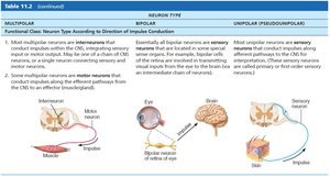
Summary Table: Structural and Functional Classes of Neurons
Neuron Type | Structural Class | Functional Class | Location |
|---|---|---|---|
Multipolar | 3+ processes (1 axon, dendrites) | Interneurons, motor neurons | CNS, motor pathways |
Bipolar | 2 processes (1 axon, 1 dendrite) | Sensory neurons | Retina, olfactory mucosa |
Unipolar | 1 process (T-like, 2 axons) | Sensory neurons | PNS, dorsal root ganglia |
Key Terms and Definitions
Nucleus (CNS): Cluster of neuron cell bodies in the CNS.
Ganglion (PNS): Cluster of neuron cell bodies in the PNS.
Nerve (PNS): Bundle of neuron processes in the PNS.
Tract (CNS): Bundle of neuron processes in the CNS.
Clinical Relevance
Certain viruses and bacterial toxins (e.g., polio, rabies, herpes simplex, tetanus toxin) can damage neural tissues by using retrograde axonal transport to reach the cell body.
Additional info:
Graded potentials are local changes in membrane potential that occur in dendrites and cell bodies, while action potentials are long-distance signals generated in axons.
Myelin increases conduction velocity by allowing saltatory conduction, where impulses jump from node to node.
