 뒤로
Gross Anatomy and Functional Organization of the Spinal Cord
스터디 가이드 - 스마트 노트
 자료에 맞춘 맞춤형 노트, 핵심 정의, 예시, 맥락을 확장해 제공합니다.
Gross Anatomy of the Spinal Cord
Structural Features
The spinal cord is a major component of the central nervous system, extending about 18 inches in length and half an inch in width. It is characterized by several anatomical landmarks and regions that facilitate its function in transmitting neural signals between the brain and the body.
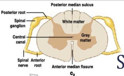
Posterior (Dorsal) Median Sulcus: A shallow longitudinal groove along the posterior surface.
Anterior (Ventral) Median Fissure: A deeper groove along the anterior surface.
Enlargements: Expanded regions of gray matter. The cervical enlargement supplies nerves to the shoulder and upper limbs, while the lumbosacral enlargement innervates the pelvis and lower limbs.
Conus Medullaris: The tapered, conical region inferior to the lumbar enlargement.
Filum Terminale: A slender strand of fibrous tissue extending from the conus medullaris.
Cauda Equina: A bundle of long anterior and posterior roots resembling a horse's tail.
Spinal Nerve Roots and Ganglia
Spinal nerves are formed by the union of anterior and posterior roots, each serving distinct functions:
Posterior Roots (Dorsal Roots): Bring sensory information into the spinal cord.
Anterior Roots (Ventral Roots): Contain axons of motor neurons that control somatic and visceral effectors.
Spinal Ganglia (Dorsal Root Ganglia): Contain cell bodies of sensory neurons.
Rootlets: Fanlike divisions of spinal nerve roots.
Spinal Nerve Branches
White Ramus: Myelinated axons.
Gray Ramus: Unmyelinated fibers innervating glands and smooth muscles.
Posterior Ramus: Sensory and motor innervation to the skin and muscles of the back.
Anterior (Ventral) Ramus: Supplies ventrolateral body surface and limbs.
Spinal Meninges
The spinal cord is protected by three specialized membranes:
Dura Mater: Tough, fibrous outermost layer.
Epidural Space: Contains areolar tissue, blood vessels, and adipose tissue.
Coccygeal Ligament: Dense cord of collagen fibers blending with filum terminale.
Arachnoid Mater: Middle layer with a delicate network of collagen and elastic fibers.
Subarachnoid Space: Filled with cerebrospinal fluid (CSF).
Pia Mater: Delicate innermost layer, firmly bound to neural tissue.
Denticulate Ligaments: Extensions from pia mater through arachnoid to dura mater.
Functional Organization of the Spinal Cord
Gray Matter
Gray matter contains neuron cell bodies and is organized into nuclei and horns:
Nuclei: Groups of neuron cell bodies.
Sensory Nuclei: Receive and relay sensory information.
Motor Nuclei: Issue motor commands to effectors.
Gray Horns: Posterior, anterior, and lateral regions of gray matter.
Gray Commissures: Bridges of gray matter connecting sides of the spinal cord.
White Matter
White matter consists of myelinated axons organized into columns and tracts:
Posterior White Columns: Between posterior horns and median sulcus.
Anterior White Columns: Between anterior horns and median fissure.
Anterior White Commissure: Interconnects anterior white columns.
Lateral White Columns: Between anterior and posterior columns.
Ascending Tracts: Carry sensory information toward the brain.
Descending Tracts: Convey motor commands from the brain.
Spinal Nerves and Peripheral Distribution
Anatomy of Spinal Nerves
Spinal nerves are composed of several layers:
Epineurium: Outermost layer of dense collagen fibers.
Fascicles: Compartments containing bundles of axons.
Perineurium: Middle layer surrounding fascicles.
Endoneurium: Innermost layer surrounding individual axons.
Peripheral Distribution
Motor Fibers: Carry motor commands to effectors.
Sensory Fibers: Carry sensory information to the CNS.
Dermatomes: Bilateral regions of skin monitored by a single pair of spinal nerves.
Nerve Plexuses
Nerve plexuses are complex networks formed by ventral rami of spinal nerves:
Cervical Plexus: C1-C5
Brachial Plexus: C5-T1
Lumbar Plexus: T12-L4
Sacral Plexus: L4-S4
Principles of Functional Organization
Neuronal Pools and Circuits
Neuronal pools are groups of interconnected neurons, organized into five main circuit types:
Divergence: Information spreads from one neuron to several neurons or pools.
Convergence: Several neurons synapse on a single postsynaptic neuron.
Serial Processing: Information relayed stepwise from one neuron to another.
Parallel Processing: Several neurons or pools process the same information simultaneously.
Reverberation: Collateral branches extend back to stimulate presynaptic neurons, forming a positive feedback loop.
Reflexes
Reflex Arc
A reflex arc is the route followed by nerve impulses to produce a reflex. The five steps in a neural reflex are:
Arrival of a stimulus and activation of a receptor
Activation of a sensory neuron
Information processing in the CNS
Activation of a motor neuron
Response by a peripheral effector
Classification of Reflexes
Development: Innate (born with) or acquired (learned motor patterns).
Nature of Response: Somatic (muscular system) or visceral (autonomic reflexes).
Complexity: Monosynaptic (direct) or polysynaptic (involving interneurons).
Processing Site: Spinal or cranial reflexes.
Monosynaptic Reflexes
The stretch reflex provides automatic regulation of skeletal muscle length. Muscle spindles, composed of intrafusal fibers, are innervated by both sensory and motor neurons. Gamma efferent motor neurons maintain sensitivity to changes in muscle length.
Postural Reflexes: Help maintain upright posture.
Polysynaptic Reflexes
Tendon Reflex: Monitors tension during muscle contraction, preventing tendon damage via Golgi tendon organs.
Withdrawal Reflexes: Move body parts away from stimulation. Includes flexor reflex and reciprocal inhibition.
Crossed Extensor Reflexes: Motor response occurs on the side opposite the stimulus, complementing withdrawal reflexes.
General Characteristics: Involve pools of interneurons, reciprocal inhibition, reverberating circuits, and coordinated responses.
Reflex Arcs
Ipsilateral Reflex Arcs: Sensory and motor responses occur on the same side.
Contralateral Reflex Arcs: Motor response occurs on the opposite side.
The Brain's Effects on Spinal Reflexes
Motor control involves multiple interacting levels. Descending tracts from the brain can activate, facilitate, or inhibit reflex motor patterns in the spinal cord by producing excitatory (EPSP) or inhibitory (IPSP) postsynaptic potentials.
Example: Reflex Modulation
A professional athlete can rapidly adjust motor patterns through acquired reflexes, while the brain can consciously override or enhance spinal reflexes during complex movements.
