 뒤로
Gross Anatomy of the Muscular System: Study Guide
스터디 가이드 - 스마트 노트
 자료에 맞춘 맞춤형 노트, 핵심 정의, 예시, 맥락을 확장해 제공합니다.
Gross Anatomy of the Muscular System
Introduction to Muscular System Anatomy
The muscular system is responsible for producing movement, maintaining posture, and stabilizing joints. Skeletal muscles attach to bones via tendons and are responsible for voluntary movements. Understanding the origin, insertion, and action of each muscle is essential for comprehending how body movements are produced and coordinated.
Origin: The fixed attachment point of a muscle, usually proximal and less movable.
Insertion: The movable attachment point, usually distal; movement occurs here when the muscle contracts.
Agonists: Muscles that produce a specific movement.
Antagonists: Muscles that oppose the action of agonists.
Types of Muscle Movements
Muscle actions are defined by the movement they produce at joints. These movements are described relative to the anatomical position:
Flexion: Decreases the angle between two bones (e.g., bending the elbow).
Extension: Increases the angle between two bones (e.g., straightening the elbow).
Hyperextension: Extension beyond the anatomical position (e.g., tilting the head backward).
Dorsiflexion: Flexing the foot upward at the ankle.
Plantarflexion: Pointing the toes downward.
Abduction: Moving a limb away from the midline (e.g., raising arms sideways).
Adduction: Moving a limb toward the midline.
Rotation: Turning a bone around its own axis (medial or lateral).
Circumduction: Circular movement combining flexion, extension, abduction, and adduction.
Inversion: Turning the sole of the foot inward.
Eversion: Turning the sole of the foot outward.
Pronation: Rotating the forearm so the palm faces posteriorly.
Supination: Rotating the forearm so the palm faces anteriorly.
Study Tips for Muscular Anatomy
Review skeletal bone anatomy to understand muscle attachments.
Use anatomical models and digital resources to locate muscles.
Visualize muscle actions using elastic bands on skeleton models.
Study in manageable sections and use repetition for memorization.
Quiz with a partner for reinforcement.
Major Muscles of the Face and Neck
Facial Muscles
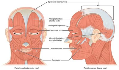
Facial muscles control expressions, mastication, and movements of the head. They are often attached to skin and other muscles rather than bones.
Muscle Name | Origin | Insertion | Action |
|---|---|---|---|
Frontalis | Epicranial aponeurosis | Fascia of facial muscles and skin above nose and eyes | Draws back scalp, wrinkles forehead, raises eyebrows |
Masseter | Maxilla and zygomatic arch | Angle and ramus of mandible | Elevates mandible (closes jaw), clenches teeth |
Temporalis | Temporal bone | Coronoid process and ramus of mandible | Elevates mandible, assists with side-to-side movement |
Orbicularis oculi | Frontal and maxilla bone | Skin of eyelids | Closes eye |
Orbicularis oris | Maxilla and mandible | Skin of upper and lower lips | Closes and protrudes lips |
Sternocleidomastoid | Sternum and clavicle | Mastoid process of temporal bone | Flexes head (both sides), rotates head to opposite side (one side) |
Muscles Involved in Ventilation
Thoracic Muscles
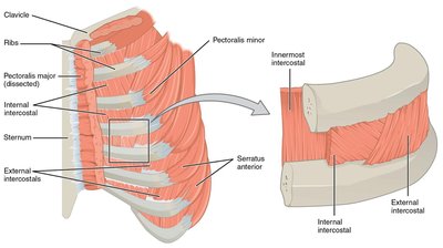
These muscles control the size of the thoracic cavity, facilitating breathing. The diaphragm is the primary muscle for inspiration, while intercostal muscles assist in both inspiration and forced expiration.
Muscle Name | Origin | Insertion | Action |
|---|---|---|---|
Diaphragm | Xiphoid process, inferior rib cage, lumbar vertebrae | Central tendon | Contraction flattens muscle, increases thoracic volume (inspiration) |
External intercostals | Inferior border of superior rib | Superior border of inferior rib | Elevates ribs during inspiration |
Internal intercostals | Superior border of inferior rib | Inferior border of superior rib | Depresses ribs during forced exhalation |
Muscles of the Abdominal Wall
Abdominal Muscles
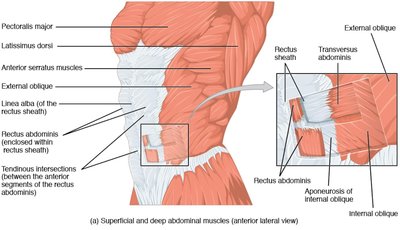
The abdominal wall consists of four main muscles: rectus abdominis (anterior), and the external oblique, internal oblique, and transversus abdominis (lateral). These muscles flex, rotate, and compress the abdominal contents.
Muscle Name | Origin | Insertion | Action |
|---|---|---|---|
Rectus abdominis | Pubic crest and symphysis | Xiphoid process, cartilage of 5th-7th ribs | Flexes and rotates vertebral column |
External oblique | 5th-12th ribs | Iliac crest, linea alba | Flexes, rotates vertebral column |
Internal oblique | Iliac crest, inguinal ligament, thoracolumbar fascia | Linea alba, cartilage of 7th-10th ribs | Flexes, rotates vertebral column |
Transversus abdominis | Iliac crest, inguinal ligament, lumbar fascia, cartilage of 5th-10th ribs | Xiphoid process, linea alba, pubis | Compresses abdominal contents |
Muscles that Act on the Pectoral (Shoulder) Girdle
Scapular Stabilizers
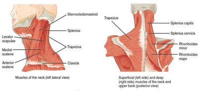
These muscles stabilize and move the scapula, aiding in arm movements. The trapezius is especially versatile due to its size and multiple fiber directions.
Muscle Name | Origin | Insertion | Action |
|---|---|---|---|
Trapezius | Occipital bone, ligamentum nuchae, spinous processes of C7 and thoracic vertebrae | Clavicle, acromion, spine of scapula | Elevates, depresses, adducts, laterally rotates scapula |
Rhomboid major | Spinous processes of T2-T5 | Medial border of scapula (inferior to spine) | Adducts, medially rotates scapula |
Rhomboid minor | Spinous processes of C7, T1 | Medial border of scapula (superior to spine) | Adducts, medially rotates scapula |
Major Muscles that Act on the Humerus
Shoulder and Arm Movers
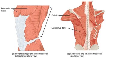
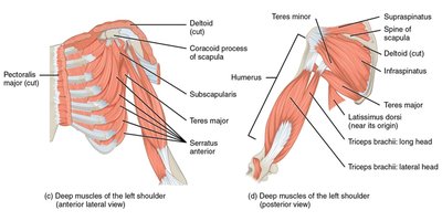
These muscles move the humerus at the shoulder joint. They include both superficial and deep muscles, with most originating on the scapula.
Muscle Name | Origin | Insertion | Action |
|---|---|---|---|
Pectoralis major | Clavicle, sternum, cartilage of 2nd-6th ribs | Greater tubercle, intertubercular sulcus of humerus | Flexes, adducts, medially rotates arm |
Latissimus dorsi | Spinous processes of T7-L5, sacrum, ilium, ribs | Intertubercular sulcus of humerus | Extends, adducts, medially rotates arm |
Deltoid | Clavicle, acromion, spine of scapula | Deltoid tuberosity of humerus | Flexes, extends, abducts, rotates arm |
Subscapularis | Subscapular fossa of scapula | Lesser tubercle of humerus | Medially rotates arm |
Supraspinatus | Supraspinous fossa of scapula | Greater tubercle of humerus | Abducts arm |
Infraspinatus | Infraspinous fossa of scapula | Greater tubercle of humerus | Abducts, laterally rotates arm |
Teres minor | Inferior lateral border of scapula | Greater tubercle of humerus | Extends, adducts, laterally rotates arm |
Teres major | Inferior angle of scapula | Intertubercular sulcus of humerus | Extends, adducts, medially rotates arm |
Muscles that Act on the Forearm
Elbow Flexors and Extensors
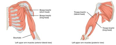
The main muscles moving the forearm at the elbow are the biceps brachii, brachialis, brachioradialis (flexors), and triceps brachii (extensor). Some also cross the shoulder joint, affecting arm movement.
Muscle Name | Origin | Insertion | Action |
|---|---|---|---|
Biceps brachii | Long head: supraglenoid tubercle; Short head: coracoid process of scapula | Radial tuberosity, bicipital aponeurosis | Flexes arm, flexes and supinates forearm |
Brachialis | Distal anterior humerus | Ulnar tuberosity, coronoid process of ulna | Flexes forearm |
Brachioradialis | Distal lateral humerus | Styloid process of radius | Flexes forearm |
Triceps brachii | Long head: infraglenoid tubercle; Lateral/medial heads: posterior humerus | Olecranon process of ulna | Extends arm and forearm |
Muscles that Act on the Upper Leg (Thigh)
Gluteal and Adductor Muscles
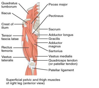
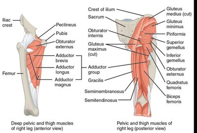
These muscles originate on the pelvis and move the thigh at the hip joint. The iliotibial tract serves as an insertion for the gluteus maximus.
Muscle Name | Origin | Insertion | Action |
|---|---|---|---|
Gluteus maximus | Iliac crest, sacrum, coccyx | Iliotibial tract, gluteal tuberosity | Extends, laterally rotates thigh |
Gluteus medius | Iliac crest | Greater trochanter of femur | Abducts, medially rotates thigh |
Adductor magnus | Inferior pubis | Linea aspera of femur | Adducts, flexes, extends, laterally rotates thigh |
Adductor longus | Anterior pubis | Linea aspera of femur | Adducts, flexes, laterally rotates thigh |
Iliopsoas | T12-L5 vertebrae, sacrum, iliac fossa | Lesser trochanter of femur | Flexes, laterally rotates thigh |
Muscles that Act on the Upper and Lower Leg
Quadriceps and Hamstrings
The quadriceps group (anterior thigh) extends the lower leg, while the hamstrings (posterior thigh) flex the lower leg and extend the thigh.
Muscle Name | Origin | Insertion | Action |
|---|---|---|---|
Rectus femoris | Anterior inferior iliac spine | Tibial tuberosity | Extends lower leg, flexes thigh |
Vastus lateralis/medialis/intermedius | Femur (various locations) | Tibial tuberosity | Extends lower leg |
Biceps femoris | Ischial tuberosity, linea aspera | Head of fibula, lateral condyle of tibia | Flexes lower leg, extends thigh |
Semitendinosus | Ischial tuberosity | Medial proximal tibia | Flexes lower leg, extends thigh |
Semimembranosus | Ischial tuberosity | Medial condyle of tibia | Flexes lower leg, extends thigh |
Other Thigh and Leg Muscles
Muscle Name | Origin | Insertion | Action |
|---|---|---|---|
Sartorius | Anterior superior iliac spine | Medial proximal tibia | Flexes lower leg, flexes and laterally rotates thigh |
Gracilis | Inferior pubis | Medial proximal tibia | Flexes lower leg, adducts thigh |
Adductor longus/magnus | Pubis/ischial tuberosity | Linea aspera, medial condyle of femur | Adducts, flexes, extends thigh, assists with lateral rotation |
Muscles that Act on the Foot and Lower Leg
Anterior and Posterior Leg Muscles
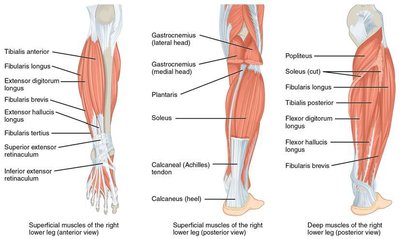
These muscles control movements of the foot and toes, as well as plantarflexion and dorsiflexion at the ankle.
Muscle Name | Origin | Insertion | Action |
|---|---|---|---|
Tibialis anterior | Lateral condyle of tibia | Medial cuneiform, 1st metatarsal | Dorsiflexes, inverts foot |
Gastrocnemius | Posterior femoral condyles | Calcaneus via Achilles tendon | Plantarflexes foot, flexes lower leg |
Soleus | Head of fibula, medial tibia | Calcaneus via Achilles tendon | Plantarflexes foot |
Additional info: For exam preparation, focus on understanding muscle actions and their anatomical relationships rather than rote memorization of origins and insertions. Practice identifying muscles on models and diagrams, and reinforce learning through active recall and repetition.
