 뒤로
L21 Hemostasis
스터디 가이드 - 스마트 노트
 자료에 맞춘 맞춤형 노트, 핵심 정의, 예시, 맥락을 확장해 제공합니다.
Blood Composition and Function
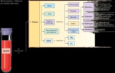
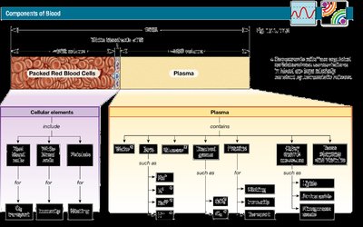
Plasma: The Fluid Matrix of Blood
Plasma is the extracellular matrix of blood, serving as the fluid component that suspends cellular elements. It is composed primarily of water, proteins, ions, organic molecules, gases, trace elements, and vitamins. Plasma is nearly identical to interstitial fluid but contains unique plasma proteins.
Water: Makes up about 92% of plasma, providing a solvent for transport.
Proteins: Constitute about 7% and include albumins (osmotic regulation), globulins (immune function), fibrinogen (clotting), and transferrin (iron transport).
Ions: Such as Na+, K+, Cl-, Ca2+, HCO3-, and H+.
Organic Molecules: Includes glucose, amino acids, lipids, and nitrogenous waste.
Gases: Oxygen (O2) and carbon dioxide (CO2).
Trace Elements & Vitamins: Essential for cellular metabolism and function.
Formed Elements: Cellular Components of Blood
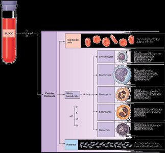
The formed elements include red blood cells (RBCs), white blood cells (WBCs), and platelets. Each plays a distinct role in physiological processes.
Red Blood Cells (Erythrocytes): Specialized for oxygen transport.
White Blood Cells (Leukocytes): Include lymphocytes, monocytes, neutrophils, eosinophils, and basophils, each with unique immune functions.
Platelets: Cell fragments essential for blood clotting and hemostasis.
Hematopoiesis: Blood Cell Production
Sites and Regulation of Hematopoiesis
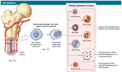
Blood cells are produced in the bone marrow through a process called hematopoiesis. Red bone marrow is active and contains hemoglobin, while yellow bone marrow is inactive and stores fat. Hematopoiesis is regulated by cytokines such as interleukins, colony-stimulating factors (CSF), thrombopoietin (TPO), and erythropoietin (EPO).
Interleukins (ILs) & CSF: Regulate leukopoiesis (WBC production).
Thrombopoietin (TPO): Controls platelet production.
Erythropoietin (EPO): Stimulates RBC production.
Bone Marrow Structure and Function
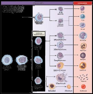
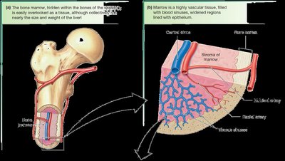
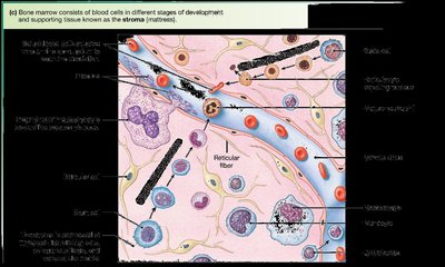
Bone marrow is a highly vascular tissue found within bones, responsible for the development of all blood cells. It contains a stroma (supportive matrix) and various cells at different stages of development.
Red Bone Marrow: Active site of hematopoiesis.
Yellow Bone Marrow: Inactive, stores adipose tissue.
Stroma: Provides structural support and microenvironment for stem cells.
Red Blood Cells (Erythrocytes)
Structure and Function
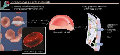
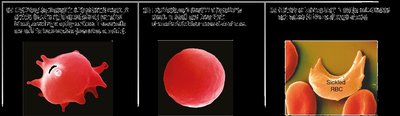
RBCs are biconcave, flexible cells specialized for oxygen transport. Their shape increases surface area for gas exchange and allows passage through capillaries.
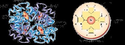
Hemoglobin: The oxygen-carrying protein, composed of four globin chains and four heme groups, each with an iron atom.
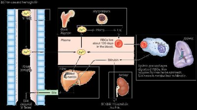
Life Span: Approximately 120 days; old RBCs are removed by the spleen.
Hemoglobin Synthesis and Iron Metabolism
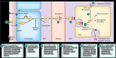
Hemoglobin synthesis requires iron, which is transported by transferrin and stored in the liver as ferritin. Iron is essential for heme production and oxygen binding.
Iron Source: Dietary intake.
Transport: Transferrin in plasma.
Storage: Ferritin in liver.
RBC Disorders
Disorders affecting RBCs can decrease oxygen transport and include anemia (low hemoglobin), hemolytic anemia, sickle cell disease, iron-deficiency anemia, and polycythemia vera.
Anemia: Reduced oxygen-carrying capacity due to low hemoglobin or RBC count.
Hemolytic Anemia: Increased RBC destruction.
Sickle Cell Disease: Abnormal hemoglobin causes RBCs to deform.
Iron-Deficiency Anemia: Insufficient iron for heme synthesis.
Causes of Anemia
Type | Cause |
|---|---|
↑ RBC Loss | Blood loss, hemolytic anemias (hereditary or acquired) |
↓ RBC Production | Defective synthesis, inadequate nutrients (iron, folic acid, B12), low erythropoietin |
Platelets and Hemostasis
Platelet Structure and Function
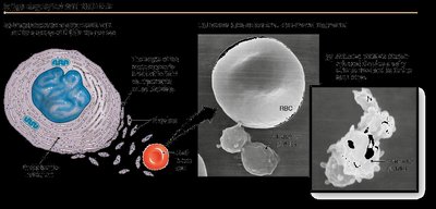
Platelets are cell fragments derived from megakaryocytes, essential for blood clotting, immunity, and inflammation. They contain mitochondria, smooth ER, and granules with cytokines and growth factors. Platelets have a lifespan of about 10 days.
Hemostasis: Prevention of Blood Loss
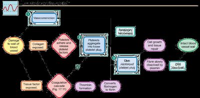
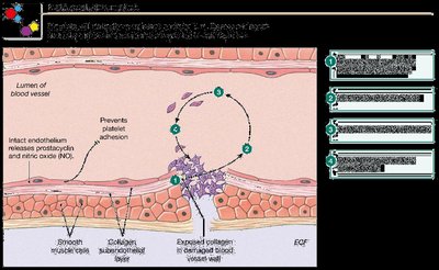
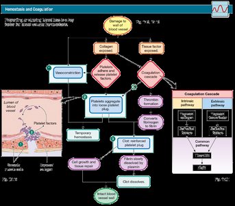
Hemostasis is the process that prevents blood loss from damaged vessels. It involves vasoconstriction, formation of a platelet plug, and activation of the coagulation cascade to form a stable clot.
Vasoconstriction: Reduces blood flow to the injury site.
Platelet Plug: Platelets adhere to exposed collagen and release factors to recruit more platelets.
Coagulation Cascade: Series of enzymatic reactions leading to fibrin formation.
Coagulation Pathways
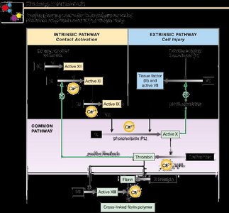
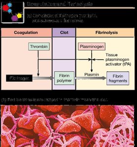
Coagulation converts a platelet plug into a clot via two pathways: intrinsic (contact activation) and extrinsic (cell injury). Both converge on the common pathway, where thrombin converts fibrinogen to fibrin, forming a stable clot.
Intrinsic Pathway: Initiated by exposure to collagen.
Extrinsic Pathway: Initiated by tissue factor from damaged cells.
Common Pathway: Thrombin formation and fibrin polymerization.
Factors Involved in Coagulation
Multiple chemical factors are involved in coagulation, including collagen, von Willebrand factor, kininogen, kallikrein, tissue factor, prothrombin, thrombin, fibrinogen, fibrin, factor XIII, Ca2+, and vitamin K.
Factor | Source | Role |
|---|---|---|
Collagen | Subendothelial ECM | Starts intrinsic pathway |
von Willebrand factor | Endothelium, megakaryocytes | Regulates factor VIII |
Tissue factor | Most cells except platelets | Starts extrinsic pathway |
Prothrombin/thrombin | Liver, plasma | Fibrin production |
Fibrinogen/fibrin | Liver, plasma | Stabilizes platelet plug |
Factor XIII | Liver, megakaryocytes | Cross-links fibrin |
Ca2+ | Plasma ions | Required for cascade |
Vitamin K | Diet | Synthesis of factors II, VII, IX, X |
Fibrinolysis: Clot Breakdown
Fibrinolysis is the process of breaking down clots. Plasminogen is activated to plasmin by tissue plasminogen activator (tPA), which dissolves fibrin and fibrinogen.
Anticoagulants and Disorders of Coagulation
Anticoagulants
Anticoagulants prevent excessive clotting by inhibiting platelet adhesion and the coagulation cascade. Endogenous anticoagulants include heparin, antithrombin III, and protein C.
Heparin: Enhances antithrombin activity.
Antithrombin III: Blocks several coagulation factors.
Protein C: Inhibits clotting factors.
Hemophilia and Coagulation Disorders
Hemophilia is a group of disorders characterized by ineffective coagulation due to defective or missing factors in the cascade. This results in prolonged bleeding and difficulty forming stable clots.
Summary of Hemostasis and Coagulation
Hemostasis involves a coordinated response to vessel injury, including vasoconstriction, platelet plug formation, coagulation, and fibrinolysis. Proper regulation is essential to prevent excessive bleeding or clotting.
Complete Blood Count (CBC) and Normal Ranges
Laboratory Values and Interpretation
A CBC provides quantitative information about blood components, including hematocrit, hemoglobin, RBC count, WBC count, differential WBC count, and platelet count. These values are essential for diagnosing blood disorders.
Test | Males | Females | Description |
|---|---|---|---|
Hematocrit | 40–54% | 37–47% | Percentage of RBCs in blood |
Hemoglobin (g/dL) | 14–17 | 12–16 | Oxygen-carrying capacity |
RBC Count (cells/mL) | 4.5–6.5 x 10⁶ | 3.9–5.6 x 10⁶ | Number of erythrocytes |
Total WBC Count (cells/mL) | 4–11 x 104 | 4–11 x 104 | Number of leukocytes |
Platelets (per µL) | 150–450 x 104 | 150–450 x 104 | Clotting ability |
Key Equations
Hematocrit Calculation
The hematocrit is calculated as:
Mean Corpuscular Volume (MCV)
Mean Corpuscular Hemoglobin (MCH)
Mean Corpuscular Hemoglobin Concentration (MCHC)
Summary Table: Blood Components
Component | Main Function |
|---|---|
Plasma | Transport, osmotic balance, clotting |
RBCs | Oxygen transport |
WBCs | Immunity |
Platelets | Clotting, hemostasis |
Summary Table: Coagulation Factors
Factor | Role |
|---|---|
Collagen | Initiates intrinsic pathway |
Tissue factor | Initiates extrinsic pathway |
Thrombin | Converts fibrinogen to fibrin |
Fibrin | Forms stable clot |
Plasmin | Dissolves fibrin (fibrinolysis) |
Additional info: Academic context was added to clarify the function and regulation of blood components, hematopoiesis, and hemostasis, as well as to provide self-contained explanations suitable for exam preparation.
