 뒤로
Influenza Virus: Structure, Pathogenesis, and Clinical Features
스터디 가이드 - 스마트 노트
 자료에 맞춘 맞춤형 노트, 핵심 정의, 예시, 맥락을 확장해 제공합니다.
Influenza Virus: Structure and Classification
Etiologic Agent and Viral Structure
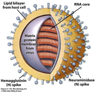
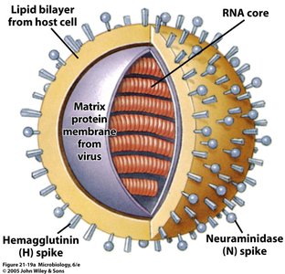
The influenza virus is a single-stranded RNA virus belonging to the Orthomyxoviridae family. It is enveloped and possesses surface spikes that play crucial roles in infection and immunity.
Etiologic Agent: Influenzavirus
Genome: Single-stranded RNA (s.s. RNA)
Envelope: Lipid bilayer derived from the host cell
Surface Spikes: Hemagglutinin (H) and Neuraminidase (N)
Matrix Protein: Provides structural integrity
Types of Influenza Virus
There are three main types of influenza virus, each with distinct epidemiological characteristics:
Influenza A: Infects humans, birds, swine, and other vertebrates; many subtypes named by H & N types (e.g., H1N1, H3N2); can cause pandemics.
Influenza B: Infects humans only; causes limited epidemics.
Influenza C: Infects humans; rarely a diagnosed problem.
Transmission and Reservoir
Reservoir and Transmission
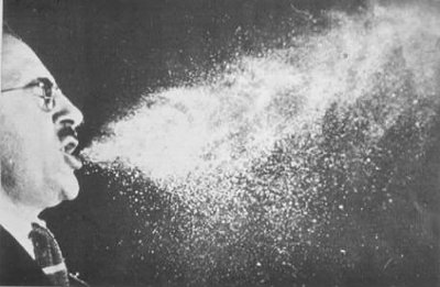
The primary reservoir for influenza viruses is infected humans. Transmission occurs mainly via aerosol droplets, which can survive outside the body for up to 24 hours.
Reservoir: Infected humans
Transmission: Aerosol droplets (coughing, sneezing)
Pathogenesis of Influenza
Initial Infection and Spread
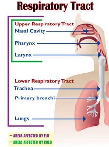
Influenza virus first invades the epithelial cells of the upper respiratory tract (URT), then may progress to the lower respiratory tract (LRT).
Target Cells: Respiratory epithelial cells
Initial Site: Upper respiratory tract (nasal cavity, pharynx, larynx)
Cellular Damage and Secondary Infection
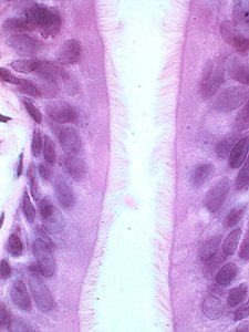
The virus destroys mucous and ciliated cells in the lower respiratory tract, compromising major host defenses and allowing secondary bacterial infections such as pneumonia.
Destruction: Mucous and ciliated cells (LRT)
Consequence: Increased risk of secondary bacterial infections
Clinical Features and Diagnosis
Symptoms and Incubation
Influenza has a short incubation period (36-48 hours) and presents with systemic and respiratory symptoms.
Incubation: 36-48 hours
Symptoms: Fever, chills, headache, malaise, muscle soreness, cough, runny nose, sore throat, fatigue, weakness (may last weeks), gastroenteritis in children
Viral Shedding: About one week
Diagnosis
Clinical Symptoms: Key for initial diagnosis
Swab Sample: Nasal swab preferred over throat swab
Rapid Test: Used for confirmation
Treatment and Prevention
Antiviral Therapy
Antiviral drugs can lessen symptoms if administered promptly (within 48 hours), but resistance may occur.
Antiviral Drugs: Effective if given early
Drug Resistance: Some viruses may develop resistance
Vaccination
Types: Killed virus (injection), live attenuated virus (nasal spray)
Recommendations: Live vaccine not recommended for certain flu seasons due to ineffectiveness
Antigenic Variation
Antigenic Drift
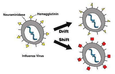
Antigenic drift refers to minor mutations in the H and N spikes, resulting in new yearly strains of influenza.
Mechanism: Point mutations in viral genome
Result: Minor changes in surface antigens
Antigenic Shift
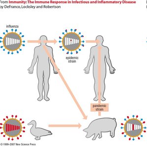
Antigenic shift involves gene reassortment, leading to dramatic changes in the virus and often preceding major epidemics.
Mechanism: Reassortment of gene segments
Result: Major changes in surface antigens
Mixing Vessels: Swine and birds can serve as hosts for reassortment

Comparison of Cold, Influenza, and Pneumonia
Clinical Differences
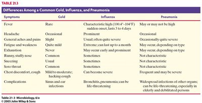
The table below summarizes the differences among common cold, influenza, and pneumonia based on symptoms and complications.
Symptoms | Cold | Influenza | Pneumonia |
|---|---|---|---|
Fever | Rare | Characteristic high (104°F–105°F), sudden onset, lasts 3–4 days | May or may not be high |
Headache | Occasional | Prominent | Occasional |
General aches and pains | Slight | Usual, often quite severe | Occasionally quite severe |
Fatigue and weakness | Quite mild | Extreme; can last up to a month | May occur, depending on type |
Exhaustion | Never | Early and prominent | May occur, depending on type |
Runny, stuffy nose | Common | Sometimes | Not characteristic |
Sneezing | Usual | Sometimes | Not characteristic |
Chest discomfort, cough | Mild to moderate; hacking cough | Can be severe | Frequent and may be severe |
Complications | Sinus and ear infections | Bronchitis, pneumonia; can be life-threatening | Widespread infections of other organs; can be life-threatening, especially in elderly and debilitated persons |
Summary
Influenza is a highly contagious respiratory virus with significant clinical and epidemiological impact. Understanding its structure, transmission, pathogenesis, and antigenic variation is essential for effective diagnosis, treatment, and prevention.
