 뒤로
뒤로Integumentary and Skeletal Systems: Structure, Function, and Clinical Correlates
스터디 가이드 - 스마트 노트
 자료에 맞춘 맞춤형 노트, 핵심 정의, 예시, 맥락을 확장해 제공합니다.
The Integumentary System
Epidermis: Structure and Types
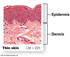
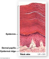
The epidermis is the outermost layer of the skin, providing a protective barrier against environmental hazards. It is composed primarily of keratinocytes and is organized into distinct strata (layers). There are two main types of skin based on the thickness of the epidermis: thin skin and thick skin.
Thin skin: Covers most of the body, contains four strata, and lacks the stratum lucidum.
Thick skin: Found on the palms of the hands and soles of the feet, contains five strata, including the stratum lucidum.
Skin Color: Determinants and Clinical Relevance
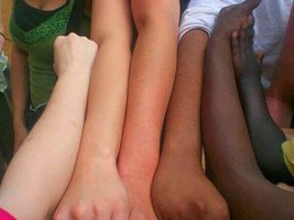
Skin color is determined by a combination of genetic and environmental factors. The main contributors include:
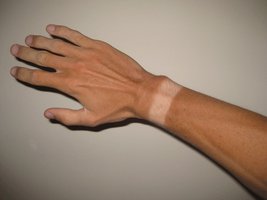
Melanin: A brown, yellow-brown, or black pigment produced by melanocytes in the stratum basale. Increased melanin production results in darker skin and provides protection against ultraviolet (UV) radiation.
Carotene: An orange-yellow pigment obtained from the diet, especially from vegetables. It can accumulate in the epidermis and subcutaneous fat.
Blood circulation: The amount of oxygenated blood in the dermal vessels can influence skin color (e.g., blushing or cyanosis).
Degree of keratinization and UV exposure can also affect pigmentation.
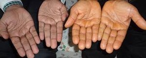
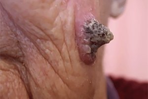
Epidermal Cancer: Types and Features
Skin cancers arise from different cell types within the epidermis. The most common types include:
Basal cell carcinoma: Originates in the stratum basale, usually due to UV-induced mutations. It rarely metastasizes and has a high survival rate.
Malignant melanoma: Arises from melanocytes, is highly aggressive, and metastasizes rapidly through the lymphatic system.
Squamous cell carcinoma: Develops from squamous cells, often due to UV exposure, and can occur in various epithelial tissues.
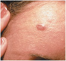
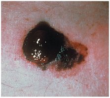
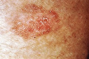
Dermis: Structure and Layers
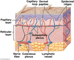
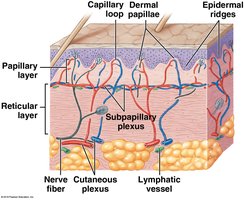
The dermis lies beneath the epidermis and provides structural support, nourishment, and sensory functions. It consists of two main layers:
Papillary layer: Composed of areolar tissue, contains capillaries, lymphatic vessels, and sensory neurons. The dermal papillae interlock with epidermal ridges, strengthening the connection between epidermis and dermis.
Reticular layer: Made of dense, irregular connective tissue, containing blood vessels, lymphatics, nerve fibers, and accessory organs such as hair follicles and glands.
The Skeletal System
Overview and Functions
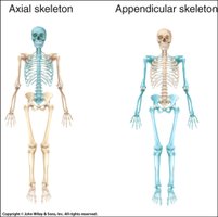
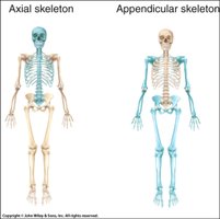
The skeletal system consists of bones, cartilages, ligaments, and other connective tissues. It is divided into the axial skeleton ( skull, thorax, vertebral column) and the appendicular skeleton (limbs and girdles). Major functions include:
Support
Mineral and lipid storage
Blood cell production
Protection of organs
Leverage for movement
Bone Classification by Shape
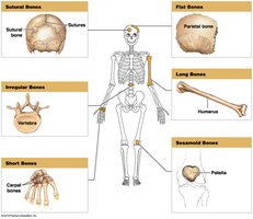
Bones are classified into six categories based on their shape and function:
Type | Description | Example |
|---|---|---|
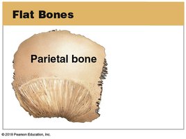
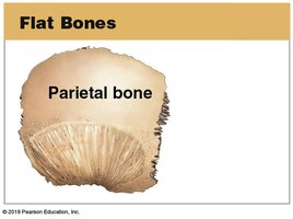
Flat bones | Thin, parallel surfaces; protect soft tissues | Parietal bone, sternum, ribs, scapulae |
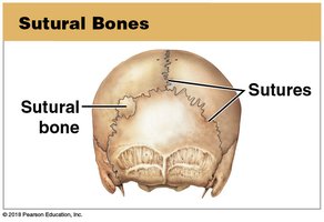
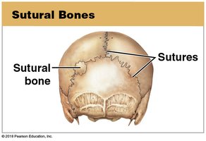
Sutural bones | Irregular bones between cranial bones | Sutures of skull |
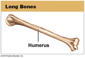
Long bones | Long and slender | Humerus, femur |
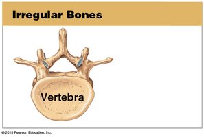
Irregular bones | Complex shapes | Vertebrae, pelvis |
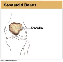
Sesamoid bones | Small, round, develop in tendons | Patella |
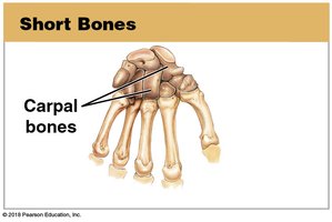
Short bones | Small and boxy | Carpals, tarsals |
Bone Markings
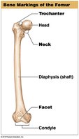
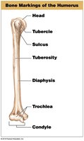
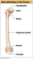
Bone markings are anatomical features on bones that serve as attachment points for muscles, passageways for nerves and blood vessels, and articulations with other bones. Key terms include:
Head: Expanded end of a bone forming part of a joint
Diaphysis: Shaft of a long bone
Neck: Narrow connection between head and diaphysis
Process: Any projection or bump
Tubercle: Small, rounded projection
Tuberosity: Broad, rough projection
Trochlea: Pulley-shaped articular process
Condyle: Smooth, rounded articular process
Trochanter: Large, rough projection
Facet: Small, flat articular surface
Long Bone Structure
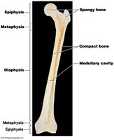
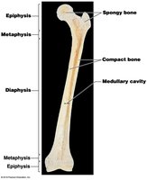
Long bones have specialized regions and coverings that support growth and function:
Epiphysis: Expanded ends, mostly spongy bone, covered by articular cartilage
Metaphysis: Connects epiphysis to diaphysis; contains the growth plate (epiphyseal plate/line)
Diaphysis: Shaft, contains the medullary (marrow) cavity
Periosteum: Outer connective tissue covering
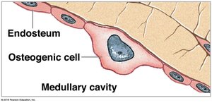
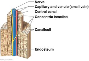
Endosteum: Inner connective tissue lining the medullary cavity
Osseous Cells and Tissues
Bone tissue contains several specialized cell types:
Osteogenic cells: Stem cells that differentiate into osteoblasts; important for bone repair.
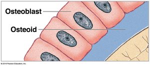
Osteoblasts: Produce new bone matrix (osteoid) and initiate ossification.
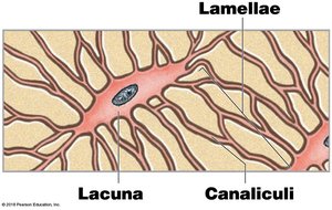
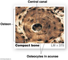
Osteocytes: Mature bone cells that maintain the matrix; reside in lacunae and communicate via canaliculi.
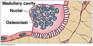
Osteoclasts: Large, multinucleated cells that resorb bone matrix, releasing minerals into the blood (osteolysis).
Compact and Spongy Bone
Bone tissue is organized into two main types:
Compact bone: Dense outer layer, composed of osteons (Haversian systems) with concentric lamellae around a central canal. Perforating canals connect osteons.
Spongy bone: Network of trabeculae with spaces for red bone marrow; lacks osteons and central canals.
Bone Growth and Remodeling
Bone growth occurs through two main processes:
Appositional growth: Increase in bone diameter by adding new layers to the surface.
Interstitial growth: Lengthening of bone at the epiphyseal plate by chondrocyte division and matrix secretion.
Bone remodeling is a continuous process involving osteoblast and osteoclast activity, regulated by hormones and mechanical stress.
Calcium Homeostasis
Calcium is the most abundant mineral in the body, with about 99% stored in bones. Blood calcium levels are tightly regulated by:
Parathyroid hormone (PTH): Increases blood calcium by stimulating osteoclasts, increasing intestinal absorption (via vitamin D), and reducing renal excretion.
Calcitonin: Decreases blood calcium by inhibiting osteoclasts, reducing intestinal absorption, and increasing renal excretion.
Disruptions in calcium homeostasis can affect muscle contraction, nerve conduction, and blood coagulation.
Bone Fractures and Healing
Fractures are classified by their characteristics:
Closed (simple): Do not break the skin.
Open (compound): Break through the skin, increasing risk of infection.
Transverse: Perpendicular to bone axis.
Spiral: Caused by twisting forces.
Compression: Occur in vertebrae under stress.
Greenstick: Incomplete break, common in children.
Comminuted: Bone shatters into fragments.
Epiphyseal: Involve the growth plate; may affect bone growth in children.
Joints (Articulations)
Functional and Structural Classification
Joints are classified by their function (degree of movement) and structure (type of tissue connecting bones):
Functional Class | Movement | Example |
|---|---|---|
Synarthrosis | No movement | Sutures of skull |
Amphiarthrosis | Little movement | Pubic symphysis |
Diarthrosis | Freely movable | Shoulder, knee |
Structural types include fibrous (sutures, gomphoses, syndesmoses), cartilaginous (synchondroses, symphyses), bony (synostoses), and synovial joints.
Synovial Joints: Structure and Function
Synovial joints are diarthrotic and allow a wide range of motion. Key components include:
Articular cartilage: Covers bone surfaces, reduces friction.
Joint (articular) capsule: Encloses the joint cavity, continuous with periosteum.
Synovial membrane: Lines the capsule, secretes synovial fluid for lubrication, nutrient distribution, and shock absorption.
Accessory structures: Ligaments, tendons, bursae, fat pads, and menisci provide support and stability.
Types of Synovial Joints and Movements
Synovial joints are classified by the shapes of their articulating surfaces and the types of movement they allow:
Plane (gliding) joint: Carpals, tarsals
Hinge joint: Elbow, knee
Pivot joint: Proximal radioulnar joint
Condylar joint: Metacarpophalangeal joints
Saddle joint: Thumb
Ball-and-socket joint: Shoulder, hip
Movements include flexion, extension, abduction, adduction, circumduction, rotation, and special movements such as opposition, inversion, eversion, protraction, retraction, elevation, and depression.
*Additional info: This guide covers the structure and function of the integumentary and skeletal systems, including clinical correlations relevant to Anatomy & Physiology students. For more detail on bone growth, hormonal regulation, and joint pathologies, refer to the corresponding textbook chapters.*
